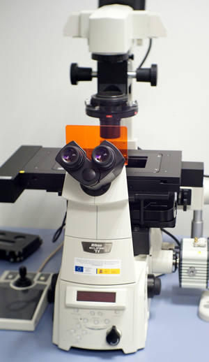
U28-E16. Nikon Eclipse Ti basic Fluorescence Microscope
Specifications:
Inverted Nikon Ti Eclipse fully motorised microscope
Hamamatsu Orca R2 monochrome 1.3 MP CCD camera
Okolab H101 stage-top incubator with temperature, humidity and CO2 control
Objective lenses:
- Plan Apo Oil immersion: 60x (1.4 NA)
- Super Plan Fluor 40x (0.6 NA) Extra Long working distance (2.8-3.6mm)
- Plan Apo 10x (0.45 NA)
Nikon Intensilight 130w Mercury Lamp
Filter sets for DAPI, GFP, TRITC
Nikon PFS (Perfect Focus System) for improved stability