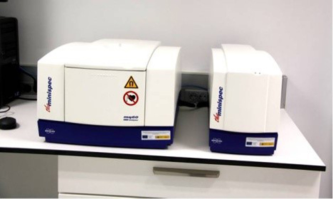
U28-E06. Bruker Minispec MQ-60
The Minispec TD-NMR is broadly used in research, development and quality control of MRI Contrast Agents. These agents are used to enhance contrast in Magnetic Resonance images between tissues that otherwise would be difficult to differentiate. Most of these contrast agents are based on Gd chelates or iron oxide nanoparticles, which produce signal contrast enhancement by affecting the magnetic relaxation properties of water molecules. The Minispec MQ-60 provides T1 and T2 measurements with high accuracy and reproducibility, and, most importantly, at clinical field, which is fundamental for translational research. Hence, the Minispec MQ-60 plays a key role in the characterization of new contrast agents for clinical use.
Specifications:
Permanent 1.4T magnetic field.
“Intelligent” 1H probe (“plug and play”) with 60 MHz operating frequency
Fast magnet warm-up and precise digital temperature regulation for ultimate results stability
Optical detector to any kind of glass, quartz and plastic samples. Specific filters for natural and fluorescent light suppression and energy saving lamps