Experimental & molecular medicine: a new article with NANBIOSIS U17
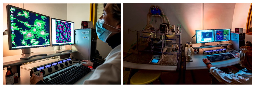
ICTS-NANBIOSIS. UNIT 17 CONFOCAL MICROSCOPY SERVICE. (CIBER-BNN. UNIVERSITY OF ALCALÁ)
The UAH research groupTranslational research of chronic diseases associated with aging and kidney disease has recently published an article in which unit NANBIOSIS unit 17 and Leica SP5 confocal microscope has had a great contribution
Located in the Support Center for Research in Medicine and Biology, Faculty of Medicine and Healthe Sciences, University of Alcalá (UAH). The Unit is equipped with a Leica TCS-SP5 confocal microscope. The confocal module is equipped with three spectral detection channels, AOBS (Acousto-optical beam splitter) and a resonant scanner system that allows analysis at high speed and resolution and makes possible the analysis of dynamic in vivo physiological processes in short periods, significantly improving the survival of living biological samples by shortening the exposure times to lasers. Includes an argon laser, a He/Ne laser, a DPSS laser diode and a violet excitation laser. The microscope is coupled to a cell incubation kit that allows multi-position time-lapse experiments. The equipment includes a workstation and four software for acquisition and analysis, which allow 3D visualizations, co-location studies, FRAP (Fluorescent Recovery after Photo-bleaching), FLIP (Fluorescent Loss in Photobleaching) and FRET (Fluorescence Resonant Energy Transfer). The equipment allows 3D characterization in detail of living cells and tissues through the use of different fluorochromes, expression and localization of molecules in 2/3D, colocalization and interaction of proteins or other types of molecules; endocytosis and intracellular transport, in situ hybridization with fluorescent probes, interaction studies between cells and materials, etc.
The unit provides researchers with a wide array of routine and specialized services as well as the latest advances in microscopy, including technical and scientific support to scientists for the study of cell/tissue biology, physiology and pathogenesis of diseases.
Article of reference
Campillo, S., Bohorquez, L., Gutiérrez-Calabrés, E., García-Ayuso, D., Miguel, V., Griera, M., Calle, Y., de Frutos, S., Rodríguez-Puyol, M., Rodríguez-Puyol, D., & Calleros, L. Indoxyl sulfate- and P-cresol-induced monocyte adhesion and migration is mediated by integrin-linked kinase-dependent podosome formation Experimental & molecular medicine, 2022, 54(3): 226–238. https://doi.org/10.1038/s12276-022-00738-8
Abstract
Cardiovascular disease is an important cause of death in patients with chronic kidney disease (CKD). Protein-bound uremic toxins, such as p-cresyl and indoxyl sulfate (IS), are poorly removed during hemodialysis, leading to vascular endothelial dysfunction and leukocyte extravasation. These processes can be related to dynamic adhesion structures called podosomes. Several studies have indicated the role of integrin-linked kinase (ILK) in the accumulation of integrin-associated proteins in podosomes. Here, we investigated the involvement of ILK and podosome formation in the adhesion and extravasation of monocytes under p-cresol (pc) and IS exposure. Incubation of THP-1 human monocyte cells with these toxins upregulated ILK kinase activity. Together, both toxins increased cell adhesion, podosome formation, extracellular matrix degradation, and migration of THP-1 cells, whereas ILK depletion with specific small interfering RNAs suppressed these processes. Interestingly, F-actin colocalized with cortactin in podosome cores, while ILK was colocalized in podosome rings under toxin stimulation. Podosome Wiskott-Aldrich syndrome protein (WASP)-interacting protein (WIP) and AKT protein depletion demonstrated that monocyte adhesion depends on podosome formation and that the ILK/AKT signaling pathway is involved in these processes. Ex vivo experiments showed that both toxins induced adhesion and podosome formation in leukocytes from wild-type mice, whereas these effects were not observed in leukocytes of conditional ILK-knockdown animals. In summary, under pc and IS stimulation, monocytes increase podosome formation and transmigratory capacity through an ILK/AKT signaling pathway-dependent mechanism, which could lead to vascular injury. Therefore, ILK could be a potential therapeutic target for the treatment of vascular damage associated with CKD.