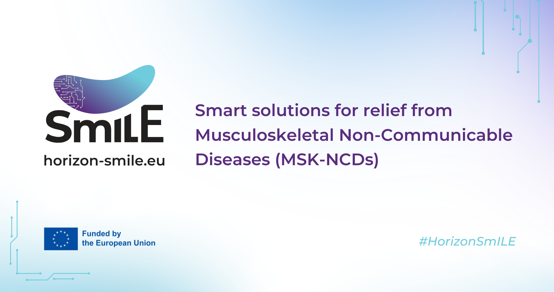
+34 620 10 75 37info@nanbiosis.com

U24. Medical imaging

U24. Medical imaging

U24-S01 Evaluation of therapies for cardiovascular disease

Evaluation of therapies for cardiovascular disease

With cardiovascular disease consistently representing a major cause of death worldwide, a platform to perform experimental studies testing the efficacy of candidate therapies for CVD is necessary.
The methodology implemented in NANBIOSIS Unit 24 for this purpose has been tested and validated in several papers (Refs below). In brief, CVD is induced in a relevant large animal model (i.e. myocardial infarction and swine) using image guided surgical techniques (i.e. percutaneous balloon occlusion of a coronary artery for a pre-determined amount of time). Once the model is established, the therapy under study is applied and the experimental subjects followed up for a fixed length of time. Clinical grade imaging (i.e. Cardiac Magnetic Resonance including delayed enhancement) and laboratory techniques are used to follow up and document the evolution of the induced CVD. Generally, image acquisition is performed at baseline and serially during the predetermined duration of the study in order to study the effect of the therapy on measurable endpoints (i.e. left ventricular ejection fraction) to document improvement.

Customer benefits

The studies are tailored to the needs of each specific candidate intervention, can be implemented with different follow-up times and can be performed under regulatory requirements, since the performing institution is Certified for Good Laboratory Practices.
Thus the Service can be provided as proof-of-concept studies, full safety and efficacy or under GLPs to meet regulatory agencies’ guidelines and assure clinical translation, so that the customers can take their therapy to a clinical trial faster and more efficiently, thanks to the full range of capabilities offered in this service.

Target customer

The offered service can be of interest to scientists from academia willing to test a possible CVD therapy, including biologicals, small companies that have a candidate molecule which is promising enough to warrant large animal trials or big pharma willing to undergo GLP studies to commercialize their product.

References

  • Aimo A et al. Colchicine added to standard therapy further reduces fibrosis in pigs with myocardial infarction. J Cardiovasc Med (Hagerstown). 2023 Nov 1;24(11):840-846. doi: 10.2459/JCM.0000000000001554. Epub 2023 Sep 29. PMID: 37773884.
  • Österberg K et al. Personalized tissue-engineered veins – long term safety, functionality and cellular transcriptome analysis in large animals. Biomater Sci. 2023 May 30;11(11):3860-3877. doi: 10.1039/d2bm02011d. PMID: 37078624.
  • Pulido M et al. Transcriptome Profile Reveals Differences between Remote and Ischemic Myocardium after Acute Myocardial Infarction in a Swine Model. Biology (Basel). 2023 Feb 21;12(3):340. doi: 10.3390/biology12030340. PMID: 36979032; PMCID: PMC10045039.
  • Blanco-Blázquez V et al Intracoronary Administration of Microencapsulated HGF in a Reperfused Myocardial Infarction Swine Model. J Cardiovasc Dev Dis. 2023 Feb 17;10(2):86. doi: 10.3390/jcdd10020086. PMID: 36826582; PMCID: PMC9960949.
  • Arenal Á et al. Effects of Cardiac Stem Cell on Postinfarction Arrhythmogenic Substrate. Int J Mol Sci. 2022 Dec 19;23(24):16211. doi: 10.3390/ijms232416211. PMID: 36555857; PMCID: PMC9781106.
  • Báez-Díaz C et al. Intrapericardial Delivery of APA-Microcapsules as Promising Stem Cell Therapy Carriers in an Experimental Acute Myocardial Infarction Model. Pharmaceutics. 2021 Nov 1;13(11):1824. doi: 10.3390/pharmaceutics13111824. PMID: 34834235; PMCID: PMC8626005.
  • Crisóstomo V et al. The epicardial delivery of cardiosphere derived cells or their extracellular vesicles is safe but of limited value in experimental infarction. Sci Rep. 2021 Nov 12;11(1):22155. doi: 10.1038/s41598-021-01728-y. PMID: 34772964; PMCID: PMC8590017.
  • Prat-Vidal C et al. Intracoronary Delivery of Porcine Cardiac Progenitor Cells Overexpressing IGF-1 and HGF in a Pig Model of Sub-Acute Myocardial Infarction. Cells. 2021 Sep 28;10(10):2571. doi: 10.3390/cells10102571. PMID: 34685551; PMCID: PMC8534140.
  • Ziani K et al. Characterization of encapsulated porcine cardiosphere-derived cells embedded in 3D alginate matrices. Int J Pharm. 2021 Apr 15;599:120454. doi: 10.1016/j.ijpharm.2021.120454. Epub 2021 Mar 5. PMID: 33676988.
  • Rossello X et al. CIBER-CLAP (CIBERCV Cardioprotection Large Animal Platform): A multicenter preclinical network for testing reproducibility in cardiovascular interventions. Sci Rep. 2019 Dec 30;9(1):20290. doi: 10.1038/s41598-019-56613-6. PMID: 31889088; PMCID: PMC6937304.
  • Crisostomo V et al. Dose-dependent improvement of cardiac function in a swine model of acute myocardial infarction after intracoronary administration of allogeneic heart-derived cells. Stem Cell Res Ther. 2019 May 31;10(1):152. doi: 10.1186/s13287-019-1237-6. PMID: 31151405; PMCID: PMC6544975.
Read More

U24-S02. Medical Imaging for Minimally Invasive Surgery (Onsite&Remote) OUTSTANDING

Medical Imaging for Minimally Invasive Surgery

Advancements in medical technologies have enabled the development and regular use of minimally invasive procedures, which could also be termed image-guided surgery. A platform that can integrate the different imaging techniques is pivotal to aid their development.
NANBIOSIS Unit 24 has medical grade imaging (i.e. Cardiac Magnetic Resonance including delayed enhancement, high performance ultrasound systems, flat panel fluoroscopes, intravascular optical coherence tomography) integrated in a surgical environment, so that these technologies are available for comprehensive follow up of interventions or new surgical approaches. Generally, image acquisition is performed at baseline and serially during the predetermined duration of the study in order to assess the effectiveness of these surgeries on measurable endpoints to document improvement.

Customer benefits

The studies are tailored to the needs of each specific candidate intervention, can be implemented with different follow-up times and can be performed under regulatory requirements, since the performing institution is Certified for Good Laboratory Practices.
Thus the Service can be provided as proof-of-concept studies, full safety and efficacy or under GLPs to meet regulatory agencies’ guidelines and assure clinical translation, so that the customers can take their therapy to a clinical trial faster and more efficiently, thanks to the full range of capabilities offered in this service.

Target customer

The offered service can be of interest to scientists from academia willing to test a possible surgery and perform serial follow up of its outcomes, early-stage medical professionals requiring training in minimally invasive surgery, late-stage surgeons that need to test an idea for an improved surgical approach, commercial companies that need to test the implantation of a device and its effect under GLP studies to commercialize their product.

References

  • Lucas-Cava V, Sánchez-Margallo FM, Dávila-Gómez L, Lima-Rodríguez JR, Sun F. Prostatic artery occlusion versus prostatic artery embolisation for the management of benign prostatic hyperplasia: early results in a canine model. Br J Radiol. 2022 Aug 1;95(1136):20220243. doi: 10.1259/bjr.20220243. Epub 2022 Jul 7. PMID: 35762334; PMCID: PMC10162044.
  • Sánchez-Margallo JA, Tas L, Moelker A, van den Dobbelsteen JJ, Sánchez-Margallo FM, Langø T, van Walsum T, van de Berg NJ. Block-matching-based registration to evaluate ultrasound visibility of percutaneous needles in liver-mimicking phantoms. Med Phys. 2021 Dec;48(12):7602-7612. doi: 10.1002/mp.15305. Epub 2021 Oct 31. PMID: 34665885; PMCID: PMC9298012.
  • Sánchez-Margallo FM, Veloso Brun M, Sánchez-Margallo JA. Identification of intra-abdominal lymphatics in canine carcasses by laparoscopic fluorescence lymphography with intradermal and intrapopliteal ICG administration. PLoS One. 2020 Nov 12;15(11):e0241992. doi: 10.1371/journal.pone.0241992. PMID: 33180854; PMCID: PMC7660503.
  • Sánchez-Peralta LF, Picón A, Sánchez-Margallo FM, Pagador JB. Unravelling the effect of data augmentation transformations in polyp segmentation. Int J Comput Assist Radiol Surg. 2020 Dec;15(12):1975-1988. doi: 10.1007/s11548-020-02262-4. Epub 2020 Sep 28. PMID: 32989680; PMCID: PMC7671995.
  • Crisostomo V et al. Dose-dependent improvement of cardiac function in a swine model of acute myocardial infarction after intracoronary administration of allogeneic heart-derived cells. Stem Cell Res Ther. 2019 May 31;10(1):152. doi: 10.1186/s13287-019-1237-6. PMID: 31151405; PMCID: PMC6544975.
Read More

U24-E01. Fluoroscopy

  • Cardiac Electrophysiology Laboratory: radiofrecuency ablation system and multicanal poligraph.
  • Fluoroscopy: BV PULSERA Philips Medical Systems
Read More

U24-E02. Digital subtraction angiography system

  • Digital Subtraction Angiography (DSA) (PHILIPS BV- Pulsera Philips Medical System, Netherlands).
  • Digital Subtraction Angiography with flat panel detector (DSA) (PHILIPS Veradius Unity
Read More

U24-E03. Ultrasound scanner

  • Ultrasound scanner (Panther Ultrasound Scanner Type 2002, B&K MEDICAL, Inc. DENMARK.
  • Ultrasound scanner (HDI5000. Philips Medical Systems, Netherlands).
Read More

U24-E04. Computed Tomography (CT) scanner

Computed Tomography (CT), Philips BRILLIANCE CT-6. The CT system includes 24 solid-state detectors that enable the acquisition of 6 simultaneous slices per 360º rotation, with a minimal slice thickness (0.6 mm) at an imaging time of 0.75 seconds per 360º for a full body scan.

These features make a high working speed possible with excellent image quality. The images can be processed in either basic or advanced mode from a workstation with the following software: Angio-CT, CINE mode, dynamic focal point reconstruction, multiplanar reconstruction (linear, curvilinear, etc), 3D multi tissue reconstruction (MIP, VR…), small volumes 3D analysis, and measurement tools for quantification (Q-CTA).

Read More

U24-E05. 1.5T Magnetic Resonance Imaging (MRI) system

Magnetic Resonance Imaging (MRI), Philips- INTERA 1.5T. The MRI equipment (Philips Intera 1.5T) enables the acquisition –using non-invasive techniques- of high definition images of any body organ at different planes, focusing on large animals as experimental models.

The availability of high precision images during research activities enables the researcher to choose correct clinical treatment. This equipment is also used in the follow up of some R&D + innovation lines; during the post operative period of experimental subjects, i.e. in activities such as cellular therapy to measure the therapeutic effect or to evaluate implanted biomaterials.

Thus, this MRI system provides a reliable diagnosis and follow up of certain diseases, allowing the evaluation of state-of-the-art health technologies and their benefits and helping to establish their clinical indications.

Read More

U24-E06. Cardiac electrophysiology lab with navigation (CARTO 3) and biotherapeutics delivery (NOGA XP) systems

Cardiac electrophysiology lab with navigation (CARTO 3) and biotherapeutics delivery (NOGA XP) systems.

Read More