Scientific leaders of the project: Prof. Nora Ventosa, Prof. Jaume Veciana and Prof. Jesús Santamaria.
Coordinator of the project: Dr Gemma Martinez and Dr. Nathaly Segovia
Industrial problem/gap covered
The conventional approaches used for the production of particulate molecular biomaterials usually follow easy preparation methods at the laboratory scale, but they fail when scaling-up to industrial level.
Description
NANBIOSIS approach offers novel synthetic strategies and experimental setup for the advanced preparation of a wide range of nanoparticles for MRIcontrast agents.
In the development of this biomedical solution, the following services are involved:
Au Magnetic /SiO2nanoshellwith a spions located inside
| Unit |
Type of NP |
Specifications of the NP (Size, composition, etc) |
Applications & reference |
| U9 |
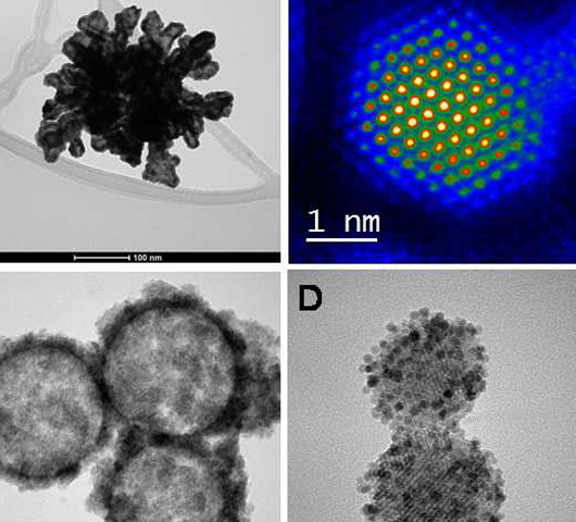
Au Magnetic /SiO2nanoshellwith a spions located inside |
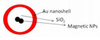
Magnetic nanoparticles in the core, covered by a shell of SiO2 with an external layer of gold, and average diameter of 100 nm |
Application: Photothermal therapy and MRI signaling (theranosticnanoparticles)
Ref:
– Nanoscale. 2014 Aug 7;6(15):9230-40. |
Hollow Au nanoshell with a spion located inside
| Unit |
Type of NP |
Specifications of the NP (Size, composition, etc) |
Applications & reference |
| U9 |
Hollow Au nanoshell with a spion located inside |
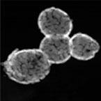
Magnetic Gold-based nanoparticles with magnetic and optical properties. Spherical shape and average diameter of 150 nm |
Application: Photothermal therapy and MRI signaling (theranostic nanoparticles)
Ref:
– Nanoscale. 2014 Aug 7;6(15):9230-40. |
Au nanorods capped with lysine
| Unit |
Type of NP |
Specifications of the NP (Size, composition, etc) |
Applications & reference |
| U9 |
Au nanorods capped with lysine |
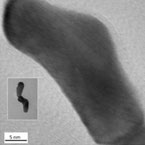
Gold-based nanorods |
Application: Contrast agents for optical coherence tomography
Ref:
– ChemCommun (Camb). 2012 Jul 7; 48(53): 6654–6656 |
Magnetic nanoparticles
| Unit |
Type of NP |
Specifications of the NP (Size, composition, etc) |
Applications & reference |
| U9 |
Magnetic nanoparticles |
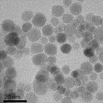
Iron-based nanoparticles with spherical shape and average diameters ranged between 6 -13 nm |
Application: MRI diagnosis (T2 contrast agents)
Ref:
J Nanopart Res (2014) 16:2292 |
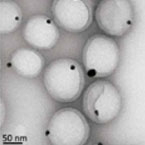
Polymeric nanoparticles with an Au nanoparticles inside
| Unit |
Type of NP |
Specifications of the NP (Size, composition, etc) |
Applications & reference |
| U9 |
Polymeric nanoparticles with an Au nanoparticles inside |
|
Application: Drug delivery and contrast agents (theranostics)
Ref:
Nanoscale, 2016,8, 6495-6506 |
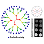
Radical dendrimers as CA for MRI
| Unit |
Type of NP |
Specifications of the NP (Size, composition, etc) |
Applications & reference |
| U6 |
Schematic representation of a Radical dendrimer |
|
Application: Contrast agents
Ref:
Organic Letters, 2013. 15(14): p. 3490-3493.
Macromolecules, 2014. 47(22): p. 7717-7724.
ChemPhysChem, 2015. 16(15): p. 3302-3307. |
 |
It gathers several laboratories, perfectly equipped, to perform the mission of this facility: the development, characterization, and large-scale production of molecular biomaterials of therapeutic or biomedical interest, with controlled micro-, nano- and supramolecular structure. One example of Key-Enabling-Technology (KET) available in this unit is a simple one-step methodology, DELOS-SUSP, based on the use of compressed fluids (CF), such as CO2, to prepare particulate materials with precise and reproducible structural characteristics at micro-, nano- and supramolecular levels (size, shape, internal structural gradients, supramolecular organization and crystalline purity).This example shows one of the singularities of this unit is that counts with CF–based plants at different scales, from mL to L, which allow process development by QbD and process scale-up.
This unit offers total characterization of radical dendrimers (structural characterization, paramagnetic properties, in vitro CA imaging properties)
– Structural characterization: NMR, UV-vis, FTIR
– Paramagnetic properties: EPR
– Relaxivity properties (MRI in vitro) |
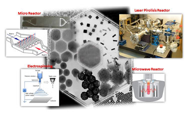 |
This unit offers novel synthetic strategies and experimental setup for the advanced preparation of a wide range of nanoparticles, including inorganic and soft nanoparticles with biomedical application.
The new methodologies include microreactors, laser pyrolysis reactor and electro spinning. Both laser pyrolysis and microreactors as belonging to the group of enabling technologies, which allow new goals in reproducibility and scale-up production of nanomaterials. As for the electro spinning, this is a new infrastructure that allows the preparation of nanowires and fibres, formed by different materials.
This unit offers total characterization of nanoparticles, nanstructures and/or colloidal suspension of nanoparticles:
– Composition (chemical analysis)
– Structure (UV-vis, NIR, XPS, FTIR, TEM, SEM)
– Suface (Gas adsorption)
– Specific properties of the nanoparticles, such as, magnetic and optical properties.
Additionally, the U9 offers study and capacity for controlled adsorption and drug delivery. |