+34 620 10 75 37info@nanbiosis.com


Custom synthesis of peptides and in vivo validation to revolutionize nanomedicine with precision drug delivery.
Scientific Leader / leaders of the project: Miriam Royo/Vanessa Diaz
Coordinator of the project: Miriam Royo/Vanessa Diaz
The delivery of therapeutically relevant amounts of drugs to specific tissues or organs is one of the major challenges in nanomedicine. Peptides have emerged as promising targeting molecules for nanomedicines due to their cost-effectiveness and ease of modification, and their conjugation to nanocarriers can specifically deliver therapeutic agents to target tissues. Selection of the appropriate targeting moiety is a critical step that should be validated in vitro and, more importantly, by in vivo biodistribution in a mouse model to assess its ability to cross some biological barriers, such as the blood-brain barrier (BBB), and its capacity to be accumulated in specific tissues or organs. The use of fluorescently labeled targeting units (peptides or small molecules) can facilitate the study of the behavior of the selected targeting unit and facilitate its selection prior to its incorporation on the nanomedicine.
In this cutting-edge, we offer the custom synthesis of fluorescently labeled peptides and the fluorescent labeling tailored to your specific nanoparticles to study their specific accumulation in the target tissues or organs. This labeling allows us to study the biodistribution of these peptides in vitro and in vivo using fluorescence or bioluminescence imaging. Different mouse models are available to determine the biodistribution of the peptides and nanomedicines, such as healthy mice or oncology mouse models. In addition, we can determine their cellular localization through histological processing.
Examples of scientific publications implementing this Cutting-Edge Biomedical Solution:
Services involved:
The projects are carried out by U3 (Synthesis of peptides Unit) and U20 (In Vivo Experimental Platform). There is a strong collaboration between these two platforms.
|
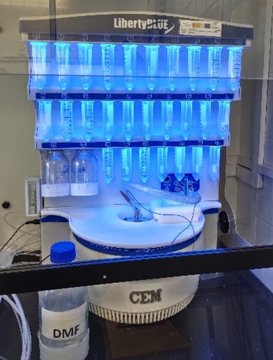
Unit 3 is coordinated by the Multivalent Systems for Nanomedicine goup at IQAC-CSIC and has the equipment necessary to provide services of synthesis of peptides at different scales (mg to g), purification, characterization, and post-synthesis modification, such as, conjugation to proteins and fluorescent. It counts with laboratories for synthesis and other to carry out modification of the peptides during and post synthesis labels. Also counts with a laboratory equipped with several preparative and analytical chromatography systems for purification and characterization. This facility benefits from the wide experience of Dr. Fernando Albericio an Dra. Miriam Royo in the design and synthesis of peptides with specific biological activity and the introduction of modifications necessary for these peptides to be bound to therapeutic nanoconjugates and other molecules, either to take advantage of the pharmacological activity of the peptide itself or to facilitate the introduction of nanoconjugates or other molecules into the cells in order to reach the therapeutic target. Its location allows the access to the rest of services of the IQAC-CSIC.. |
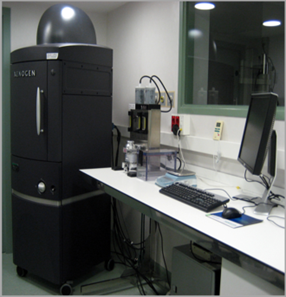 |
Located at the Valld’Hebron Research Institute (VHIR) in Barcelona, Unit 20 In vivo Experimental Platform has three different sections, a Molecular Imaging section for in vivo, ex vivo and in vitro imaging studies (fluorescence, bioluminescence and X-rays), a preclinical animal model section and a preclinical histology section. All three sections are included within the Functional Validation & Preclinical Research (FVPR) of CIBBIM-Nanomedicine. This unit is led by Dr. Ibane Abasolo (Head of FVPR). Its mission is to offer products and services for the preclinical proof-of-concept validation of therapeutic compounds and biomarkers with potential clinical applications. One of the distinguishing features of this unit is that basic preclinical studies including toxicology, histopathology and efficacy treatments are complemented with non-invasive optical imaging technologies. In vivo bioluminescence and fluorescence imaging allows monitoring of living mice longitudinally, offering real time insight into treatment efficacy, whole-body biodistribution and target mechanisms. Although in Spain there are other institutions with similar equipment, this is the only case in which imaging services can be combined with specifically devoted animal models. |