+34 620 10 75 37info@nanbiosis.com


This service is dedicated to the custom synthesis of oligonucleotide conjugates including oligonucleotides carrying lipids, amino acids, peptides or carbohydrates. In addition, they may include other modifications such as phosphorothioate linkages, 2’-O-methyl-RNA, 2’-O-MOE-RNA, 2′-F-RNA, Locked nucleic acids (LNA), modified nucleotides. The oligonucleotides will be prepared in 1 micromol scale. In most cases, the preparation of the conjugates will require the preparation of oligonucleotides carrying reactive groups such as; amino or thiol groups, which will be subjected to a post-synthetic modification. Purification and characterization will include reversed-phase HPLC analysis and mass spectrometry (MALDI-TOF).
Modification of oligonucleotides during and post synthesis to meet user requirements:
Customer benefits
The service benefits from the 40-years’ experience of the researchers participating in the service, with hundreds of scientific communications on improving the methodology used for the synthesis of modified oligonucleotides. This includes fatty acid derivatives, amino acids, cell penetrating peptides and carbohydrates not available from other services. In addition, the service can develop customized solutions for molecules not addressed in the bibliography.
Target customer
The primary audience are research groups and companies working on gene therapy and gene inhibition in the early stages of preclinical development.
References
1) “Aptamer-peptide conjugates as a new strategy to modulate human α-thrombin binding affinity”. Aviñó, A. et al. Biochim. Biophys. Acta (General subjects), 1863, 1610-1630 (2019).
2) “Synthesis of oligonucleotides carrying amino-lipid groups at the 3’-end for RNA interference studies”. Grijalvo, S. et al. J. Org. Chem., 75, 6806-6813 (2010).
3) “Synthesis and evaluation of 3’ oleyl-oligonucleotide conjugates as potential cellular uptake enhancers”. Navarro, N. et al. SYNLETT, in press (2024). doi: 10.1055/s-0042-1751528.
This service is dedicated to the custom synthesis of modified nucleotides such as nucleoside monophosphates or triphosphates as well as special phosphoramidites or functionalized solid supports functionalized with small molecules resistant to ammonia deprotection for the preparation of oligonucleotide conjugates.
Special nucleotides for oligonucleotide synthesis
For those services identified as outstanding, at least 20% of their capacity is open under competitive access. See Annex 1 of ACCESS PROTOCOL (provided by Nanbiosis) for details on % of openness for each service
Customer benefits
The service benefits from the 40-years’ experience of the researchers participating in the service with hundreds of scientific communications on improving the methodology used for the synthesis of modified oligonucleotides. This includes a range of modified nucleotides and terminal modifications not available in other services. In addition, the service can develop customized solutions for molecules not covered in the bibliography.
Target customer
The primary audience are research groups and companies involved in gene therapy, gene silencing and the development of nucleic acid-based diagnostic tools.
References
1) “Oligonucleotides containing 1’-aminomethyl or 1’-mercaptomethyl-2’-deoxy-D-ribofuranoses: Synthesis, purification, characterization and conjugation with fluorophores and lipids”. Martín-Nieves, V. et al. Bioconjugate Chem, 32, 350-366 (2021).
2) “Efficient bioactive oligonucleotide-protein conjugation for cell-targeted cancer therapy”. Aviñó, A. et al. Chemistry Open, 8, 382-387 (2019).
3) “Thioctic acid derivatives as building blocks to incorporate DNA oligonucleotides onto gold nanoparticles”. Pérez-Rentero, S. et al. Molecules, 19, 10495-10523 (2014).
This service is dedicated to the custom synthesis of modified oligonucleotides including phosphorothioate linkages, 2’-O-methyl-RNA, 2’-O-MOE-RNA, 2′-F-RNA, Locked nucleic acids (LNA), modified nucleotides and several others. The oligonucleotides will be prepared on a 1 micromol scale. Purification and characterization will include reversed-phase HPLC analysis and mass spectrometry (MALDI-TOF).
Synthesis of oligonucleotides at various different scales (100 microg to 5 mg) and purification using HPLC and/or desalting.
Customer benefits
The service benefits from the 40-years’ experience of the researchers involved in the service, with hundreds of scientific communications on improving the methodology used for the synthesis of modified oligonucleotides. This expertise includes a range of modified nucleotides and terminal modifications that are not available in other services. In addition, the service can develop customized solutions for molecules not covered in the bibliography.
Target customer
The primary audience are research groups and companies involved in mutagenesis, gene therapy, gene inhibition, the development of nucleic acid-based diagnostic tools, or the study of nucleic acid structure and nucleic acid-protein interaction.
References
1) “Detection of SARS-CoV-2 virus by Triplex Enhanced Nucleic Acid Detection Assay (TENADA)”, Aviñó, A., et al. Int. J. Mol. Sci., 23, 15258 (2022).
2) “Properties of parallel tetramolecular G-quadruplex carrying N-acetylgalactosamine as potential enhancers for oligonucleotide delivery to hepatocytes”. Clua, A. et al. Molecules, 27, 3944, (2022).
3) “Chemical modifications in nucleic acids for therapeutic and diagnostic applications”. Fàbrega, C., Aviñó, A., Eritja, R. The Chemical Record, 22, e202100270 (2022).
This service is responsible for conducting regulatory studies for the pharmaceutical industry and interested companies. The safety and efficacy studies are carried out using small and large animal models for the different organic systems, also including animal models of different pathologies.
Customer benefits
These studies are carried out under strict quality regulations, certified with ISO-9001 and Good Laboratory Practices (GLP), quality standards that allow the production of high-precision results.
Therefore, safety and efficacy studies in animal models can be carried out in compliance with the strict guidelines of regulatory agencies, ensuring the reliability and traceability of all results and tests carried out in their different services.
Target customer
The services offered in this unit may be of interest to different companies and laboratories that work within the pharmaceutical industry. Companies whose objective is to test possible candidates for molecules, drugs or medical devices in animal models of specific pathologies.
References
High content screening (HCS) attempts to bridge the gap between microscopy and cytometry, providing both large quantities of statically useful and phenotypically rich data. HCS combines automated microscopy with customizable algorithms able to detect and quantify many different types of phenotypic information in both fixed and living cultured cells. The Perkin Elmer Operetta HCS system combines automated imaging with advanced Harmony HCS software, which contains many common algorithms such as nuclei and cell detection, as well as the ability to automatically detect complex phenotypes through machine learning.
Customer benefits
We have SOPs and ISO9001 certification. We also have specialist technicians for the use of the equipment.
This service is essential to:
Target customer
Any company or research group interested in:
References
Conventional widefield fluorescence microscopy is still the best choice for many applications. The CCD and CMOS-based sensors used for conventional microscopy are typically much more sensitive than the photomultiplier tubes used in confocal microscopes and flow cytometers. As the camera captures the whole field of view at the same time, it also allows for faster imaging in many cases. Examples where conventional microscopy may be advantageous include the visualization of individual molecules, receptors, or small organisms such as bacteria and yeast.
Customer benefits
We have SOPs and ISO9001 certification. We also have specialist technicians for the use of the equipment.
This service is essential to:
Target customer
Any company or research group interested in:
References
Carrillo P, Bernal M, Téllez-Quijorna C, Marrero AD, Vidal I, Castilla L, Caro C, Domínguez A, García-Martín ML, Quesada AR, Medina MA, Martínez-Poveda B. The synthetic molecule stauprimide impairs cell growth and migration in triple-negative breast cancer. Biomed Pharmacother. 2023 Feb;158:114070. doi: 10.1016/j.biopha.2022.114070. Epub 2022 Dec 14. PMID: 36526536.
Total Internal Reflection Fluorescence (or TIRF) is a powerful technique that combines the sensitivity of conventional fluorescence with selective illumination to improve the contrast of features very close to the sample coverslip. TIRF is often used for studies related to membrane dynamics, receptor-ligand interactions, and vesicular transport.
The intrinsic diffraction of light has historically made it difficult to use fluorescence to distinguish structures closer than 200nm apart. Super-resolution microscopy refers to techniques that selectively activate fluorescent molecules to map their position with up to 10 times more accuracy than conventional fluorescent microscopy. The Nikon N-Storm system is capable of localizing molecules with a resolution of up to 20nm and is compatible with all most current localization protocols and fluorophores (including Alexafluor 647 and Atto488).
Customer benefits
We have SOPs and ISO9001 certification. We also have specialist technicians for the use of the equipment.
This service is essential to:
Target customer
Any company or research group interested in:
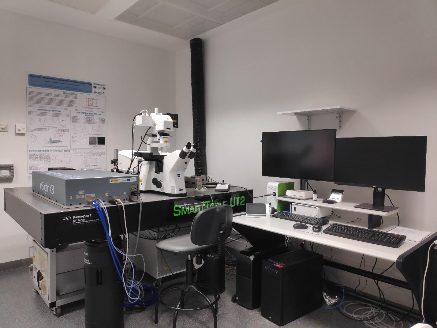
Multi- or two-photon (2P) microscopy takes advantages of the near simultaneous absorption of two or more photons which act to excite a fluorescent molecule with the combined energy of all the photons. In practice, this means that lower energy infrared (IR) light can be used to see fluorescent molecules that are normally excited by high energy ultraviolet and visible wavelength. As IR light is less susceptible to diffusion and absorption, we can visualise fluorescent molecules at greater depths than conventional microscopy. Infrared light also tends to beless damaging to live tissues than UV or blue excitation, making it ideal for timelapse imaging of model organisms or tissue explants.
Customer benefits
We have SOPs and ISO9001 certification. We also have specialist technicians for the use of the equipment.
This service is essential to:
Target customer
Any company or research group interested in:
References