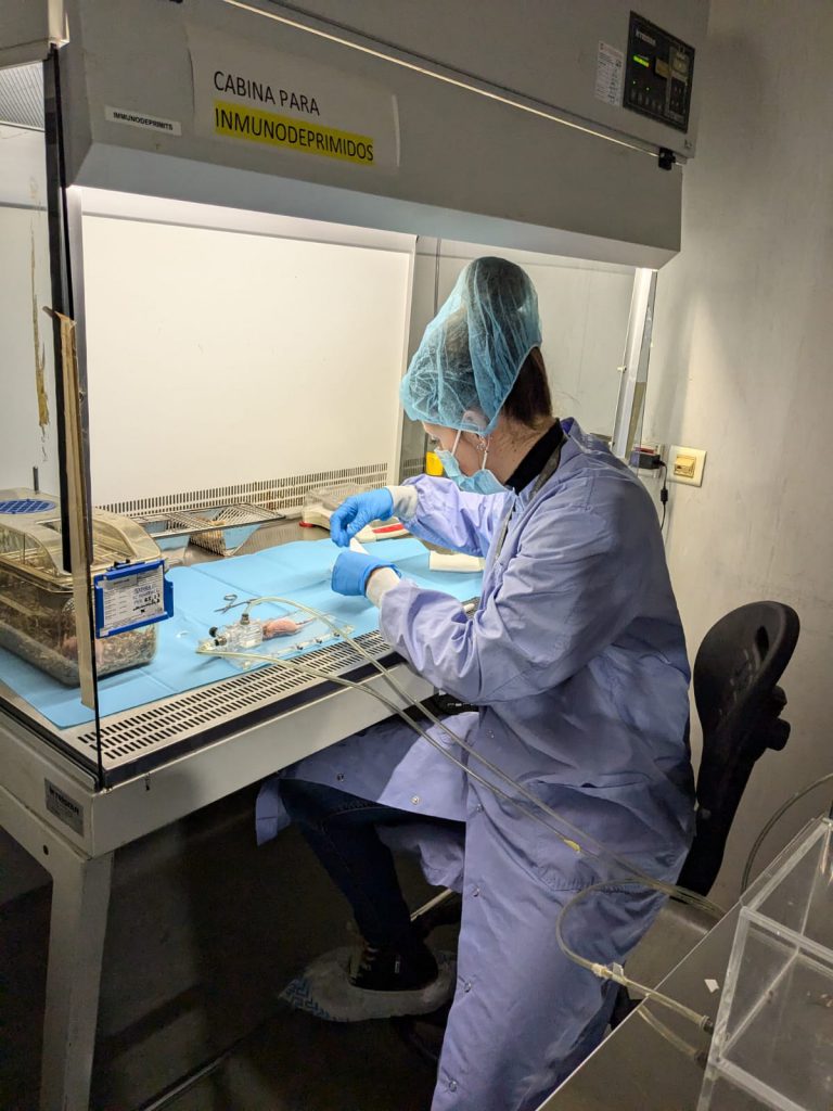
U22-S01. Animal Housing (Remote) OUTSTANDING
It houses different animal species, whether small or big (rats, mice, rabbits, dogs, pigs, sheep, etc), in compliance with the physical and legal conditions for experimental research.
The aim of this service is to evaluate the in vitro toxicity of compounds in different types of normal human cells in which we determine the effect using different types of assays including MTT, LDH release and caspase activation. The cellular models that we offer include cells from kidney, liver, bone marrow stroma or peripheral blood. Additionally, we are offering immunology assays including detection of NO production and chemotaxis and phagocytosis assays.
Customer benefits
The costumer will benefit from more than 20 years of experience of the researchers involved in the service that have performed the in vitro evaluation of the toxicity of different nanoparticles and other types of compounds. We offer also the possibility of setting up new in vitro assays or to offer other cellular models that could be of interest for the costumer. Moreover, the conditions of the assays are flexible and will be adapted to the needs of each specific compound.
Target customer
The offered service can be of interest to research groups of academia or companies willing to test the toxicity of any compound in vitro.
References
The aim of this service is to evaluate the in vivo biodistribution, toxicity and therapeutic index of nanoparticles and drug-loaded nanoparticles. Thus, we offer different mouse models in which we can determine the biodistribution of nanoparticles, with or without loaded drug, in normal and tumor tissues. Moreover, we can also determine the toxicity of the compounds and their antitumor activity in different tumor models. In the service we have subcutaneous and orthotopic/disseminated models of lymphoma, leukemia and colorectal, endometrium, head and neck carcinomas. Moreover, we have also available PDX models of colorectal and endometrium cancer. We can also offer the possibility to set up new animal models of other tumor types.
Customer benefits
The costumer will benefit from the experience of the researchers involved in the service that have performed the in vivo evaluation of a high number of diverse nanoparticles. The service has also high expertise in developing new cancer animal models. Thus, we offer also the possibility of setting up new models of any cancer type that fit with the needs of the costumer. Moreover, the conditions of the assays are flexible and will be adapted to the need of each specific compound.
Target customer
The offered service can be of interest to research groups of academia or companies willing to test the biodistribution, toxicity or antitumor activity of nanoparticles or drug-loaded nanoparticles in vivo.
References
The Confocal Microscopy Service offers the possibility of making optical section of samples and enabling their three-dimensional reconstruction, which allows their visualization in high resolution with exceptional clarity and detail.
The unit is equipped with a Leica TCS-SP5 confocal microscope. The confocal module is equipped with three spectral detection channels, AOBS (Acousto-optical beam splitter) and a resonant scanner system that allows analysis at high speed and resolution and makes possible the analysis of dynamic in vivo physiological processes in short periods of time, significantly improving the survival of living biological samples by shortening the exposure times to lasers. Includes 1 argon laser, 1 He/Ne laser, 1 DPSS laser diode and 1 violet excitation laser. The microscope is coupled to a cell incubation kit that allows multi-position time lapse experiments. The equipment includes a workstation and four software for acquisition and analysis, which allow 3D visualizations, co-location studies, FRAP (Fluorescent Recovery after Photo-bleaching), FLIP (Fluorescent Loss in Photobleaching) and FRET (Fluorescence Resonant Energy Transfer). The equipment allows 3D characterization in detail of living cells and tissues through the use of different fluorochromes, expression and localization of molecules in 2/3D, colocalization and interaction of proteins or other types of molecules; endocytosis and intracellular transport, in situ hybridization with fluorescent probes, interaction studies between cells and materials, etc.
The unit provides researchers with a wide array of routine and specialized services as well as the latest advances in microscopy, including technical and scientific support to scientists for the study of cell/tissue biology, physiology and pathogenesis of diseases. Overall, the Confocal Microscopy Service serves as a valuable tool to advance scientific research and understand complex biological systems at the cellular and molecular level.
Customer benefits
Confocal microscopy is useful in biomedical science, cell and molecular biology and physiology for visualizing subcellular organelles, cellular interactions, disease mechanisms, co-localization studies, analysis of gene expression or in vivo and real-time experiments using markers or fluorescent fusion proteins.
Also, in tissue engineering and materials science, confocal microscopy enables surface morphology characterization, and material properties.
Confocal Microscopy Service offers specific advantages to customers by providing high-resolution imaging and adding value through access to advanced instrumentation and expertise. It becomes essential for organizations involved in Research and Development across various sectors, enabling them to advance scientific understanding, accelerate innovation, and drive discoveries that have social impact.
Target customer
The Confocal Microscopy Service becomes essential for organizations involved in Research and Development in various scenarios:
References
Temporarily OUT OF ORDER
Use of the system to measure the thickness of layers, and the composition, porosity and roughness of materials on a surface.
Tests in real time to measure:
·· Molecule-molecule interactions: Protein-protein, Receptor-ligand, Antibody-antigen, Biomaterial-molecule interactions, Biomaterial-cell interactions.
·· Microbial growth.
·· Cell metabolism.
Experiments of stability, growth,
etc, perfusion and dilution
Surface analysis of organic and inorganic materials (mass spectrum), map of chemical elements present in the surface of the sample (image), profile analysis shows sample analysis in depth.
Magnetometry on nanoparticles in solid form, ultra thin films, powders, liquids and even slurries:
Measurements of magnetic properties of materials:
Geophysical Research:
Biomedical Research:
Measurement of the relaxation times in aqueous solutions and biological samples containing superparamagnetic nanoparticles as contrast agents for MR images. For T1 in the continuous range 10 kHz to 80MHz; for T2 in the range 10 MHz to 80MHz.
Characterization of contrast agents for MR imaging, ascertaining their relaxivity and the dominant effect.
NMR relaxometry technique is an important analytical tool for NMR research and material characterization in both industrial and academic environments and has been successfully applied in a wide range of fields:
– Pharmaceutical Applications:
– Polymers Applications:
– Oil, gas and petroleum applications: