+34 9340006100 -ext. 437807-info@nanbiosis.com

U26. NMR: Biomedical Applications II

U26. NMR: Biomedical Applications II

U26-S06. Biomedical and metabolomic imaging (On-site)


Description:

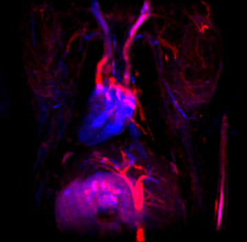
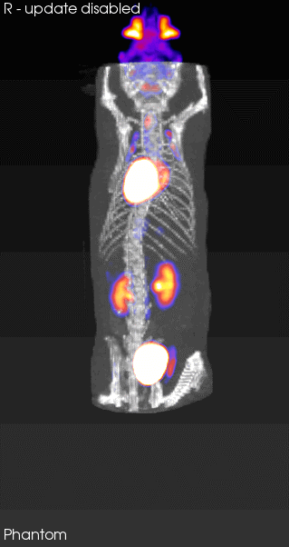
The service includes the housing of mice or rats to be able to carry out longitudinal studies, the administration, if applicable, of contrast agents, or radiopharmaceuticals for PET. Acquisition of both T1-weighted and T2-weighted images, as well as colocalized with the PET image. Volume measurement.

Applications

In addition to the typical studies in 3T preclinical imaging equipment, the possibility of varying the angle of the bed with respect to the horizontal makes it especially interesting for vascular and plant studies.

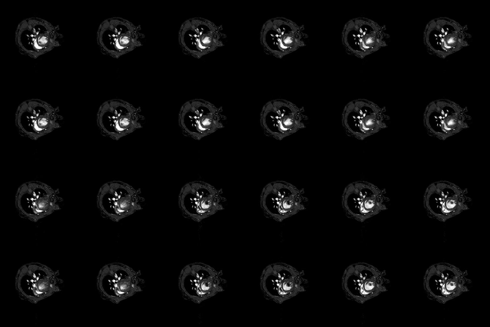
  • Qualitative and quantitative studies (relaxation times T1 and T2 and even making maps) at the brain level and other organs.Cardiological sequences: diffusion, determination of blood volume…
  • Anatomical and functional studies of different organs (for example: renal, abdominal, liver…).
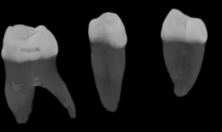
  • Bone-level imaging, distinguish between bone material from soft tissue.
  • Fat suppression techniques.
  • Oncological studies to detect and locate tumors and volume calculation.
  • Carry out studies on fruits and vegetables (grapes, kiwi, tomato, seeds…).
  • Perform studies with/without contrast (Gadolinium) and see the differences between the 2 images.
  • Thanks to the possibility of rotating the equipment in a vertical position, images of plants can be obtained (because by their nature, they have to be in a vertical orientation, such as a stem). Ex-vivo studies can also be carried out vertically and in this way we maintain the integrity of the organ (eg heart).
Read More

U26-E07. MRS*DRYMAG3017-Flexiscan

Equipment Features:

Description: 3T preclinical imaging equipment with PET ClipOn attached.

Technical specifications: Imaging equipment with superconducting magnet, capable of varying the angle from 0 to 90º, without cryogenics of 3 Teslas; mouth diameter of 170mm; 600 mT/m gradients with 5% linearity; 30 mm DSV for Homogeneity and 0.05 ppm/hour for stability; 350 Kg of total weight.

Applications:

Acquisition and viewing of in vivo images of mice and rats as well as other beings or materials of similar size.

  • Qualitative and quantitative studies (relaxation times T1 and T2 and even making maps) at the brain level and other organs.
  • Cardiological sequences: diffusion, determination of blood volume…
    Anatomical and functional studies of different organs (for example: renal, abdominal, liver…).
  • Bone-level imaging, distinguish between bone material from soft tissue.
    Fat suppression techniques.
  • Oncological studies to detect and locate tumors and volume calculation.
  • Carry out studies on fruits and vegetables (grapes, kiwi, tomato, seeds…).
  • Perform studies with/without contrast (Gadolinium) and see the differences between the 2 images.
  • Thanks to the possibility of rotating the equipment in a vertical position, images of plants can be obtained (because by their nature, they have to be in a vertical orientation, such as a stem).
  • Ex-vivo studies can also be carried out vertically and in this way we maintain the integrity of the organ (eg heart).
Read More

U26-S05. Training Advice and assistance (On-site&Remote)

Training Advice and assistance (On-site&Remote)

Read More

U26-S04. In vivo NMR of small animals(zebrafish,Drosophila (Remote) OUTSTANDING

Read More

U26-S03. Metabolomic Studies (Remote) OUTSTANDING

Metabolomic Studies

Read More

U26-S02. NMR experiments in solid state (Remote) OUTSTANDING

NMR experiments in solid state

Read More

U26-S01. Acquisition of mono and bidimensional spectra (On-site&Remote) OUTSTANDING

Acquisition of mono and bidimensional spectra

Read More

U26-E06. Gilson 215 liquid handler for automated sample preparation of liquid samples

Read More

U26-E04. Bruker DPX300 spectrometer(7 T) with standard entrance, and two RF channels.

Accessories:

  • 5mm QNP (1H, 13C, 31P and 19F), with pneumatic accessory for automatic nucleus change and field gradients along the z axis.
  • BACS autosampler for 60 samples.
Read More

U26-E02. -NMR microscopy system for imaging Bruker 14T

  • Functional and metabolic studies in cells, tissues and animal models. Coils 2mm, 5mm and 10mm. Image of 1H, 13C and 31P.
  • Microcamera for its use in the NM R equipment for:
  • The simultaneous determination of structural images (dimensions and sections of 50 μm and less)
  • Metabolic images (voxel sizes < 200 μm) in model systems.
Read More