


U18-E08. Electrophoresis systems
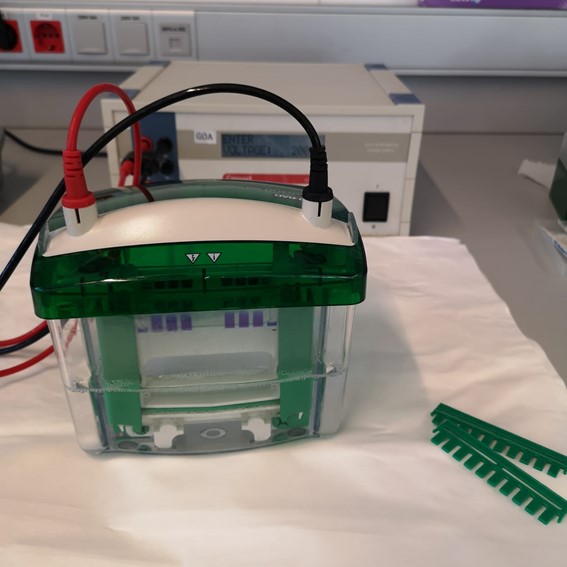
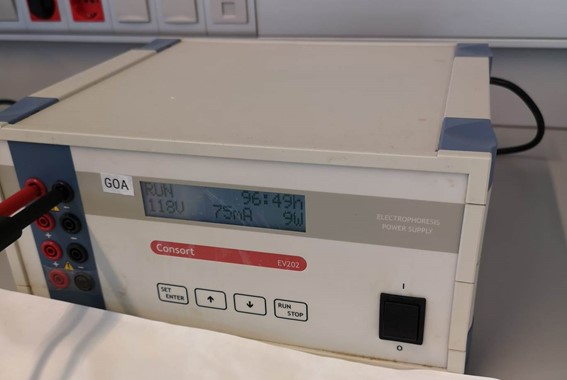
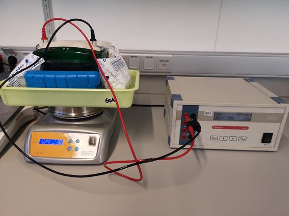
Electrophoresis of cell extracts is performed to evaluate protein expression by Western Blot assays.

Electrophoresis of cell extracts is performed to evaluate protein expression by Western Blot assays.
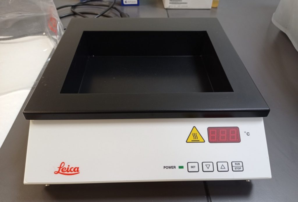
These equipments (HistoCore BIOCUT Leica microtome, a cooling plate and a bath) are used for cutting paraffin blocks of histological samples in order to perform hematoxilin&eosin staining or immunohistochemistry analyses.
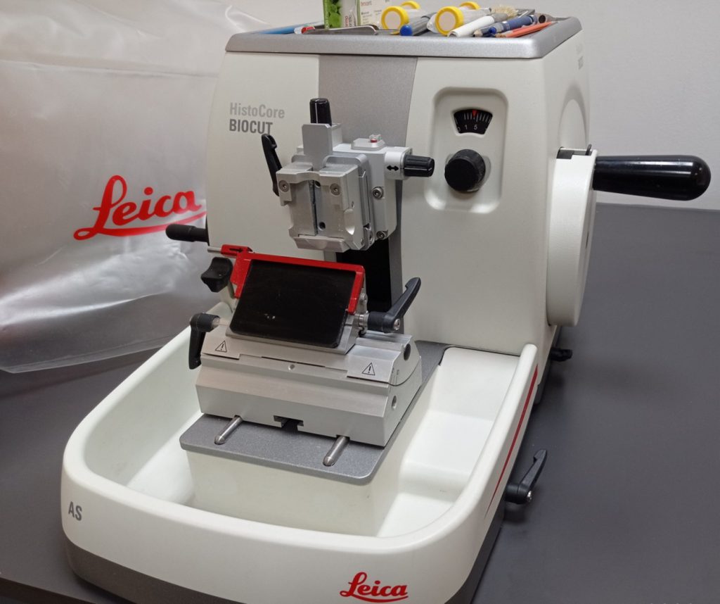
Microtome
Bath
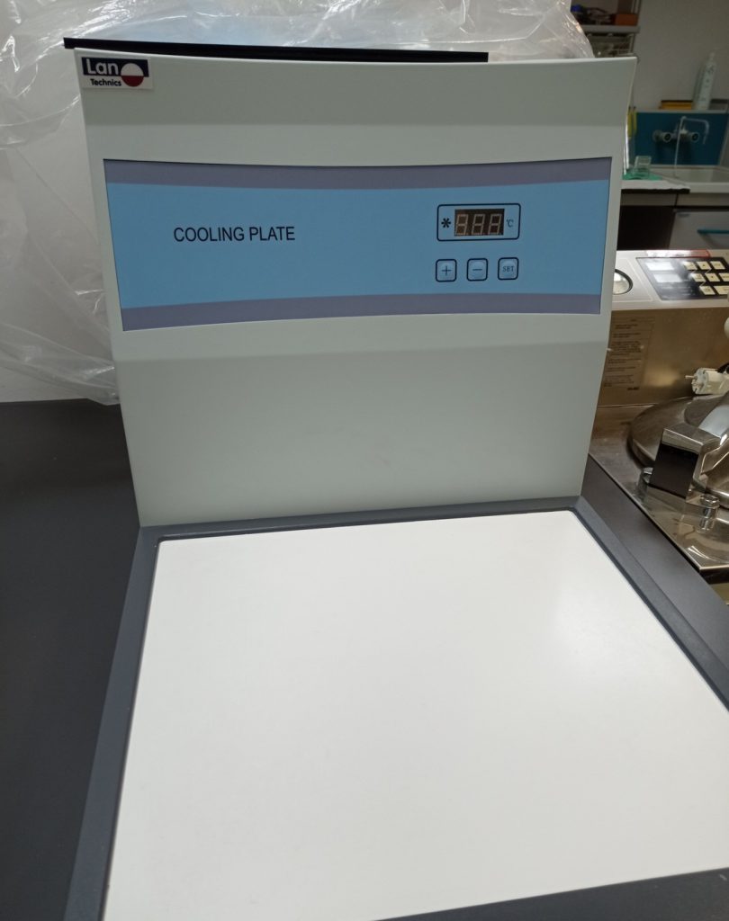
Cooling plate
Autoclaves for sterilization of buffers, media or material (tips, tubes, eppendorfs,…). Refrigerators, freezers (-20ºC and -80ºC) and liquid nitrogen tanks for the preservation of reagents, samples and cells.
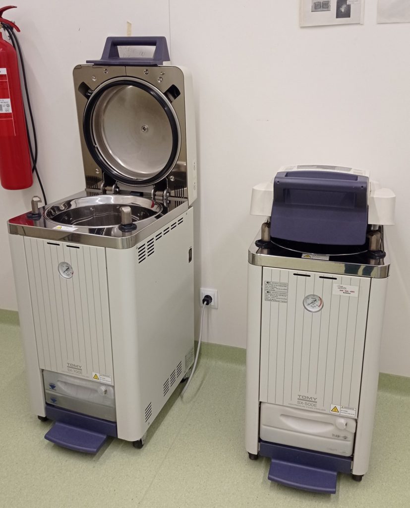
Autoclaves
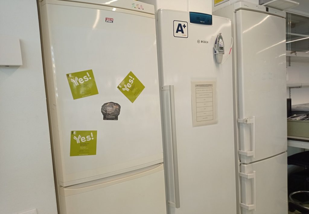
Refrigerators and -20ºC freezers
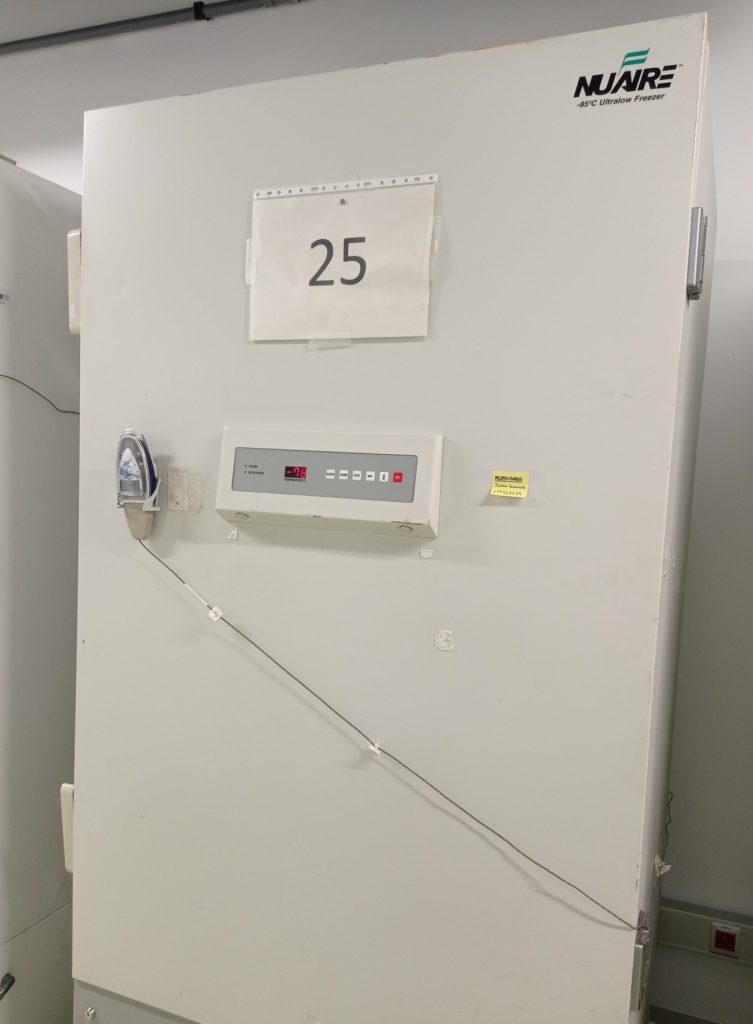
Freezers (-80ºC)
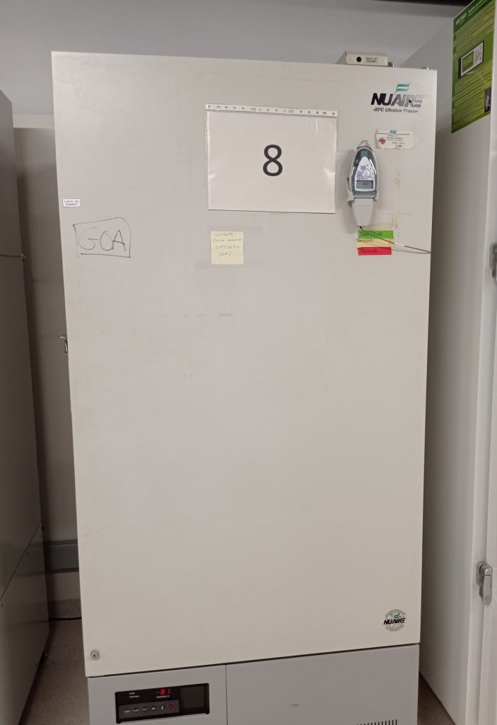
Freezers (-80ºC)
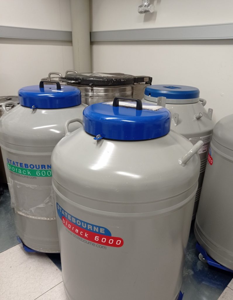
Nitrogen liquid tanks
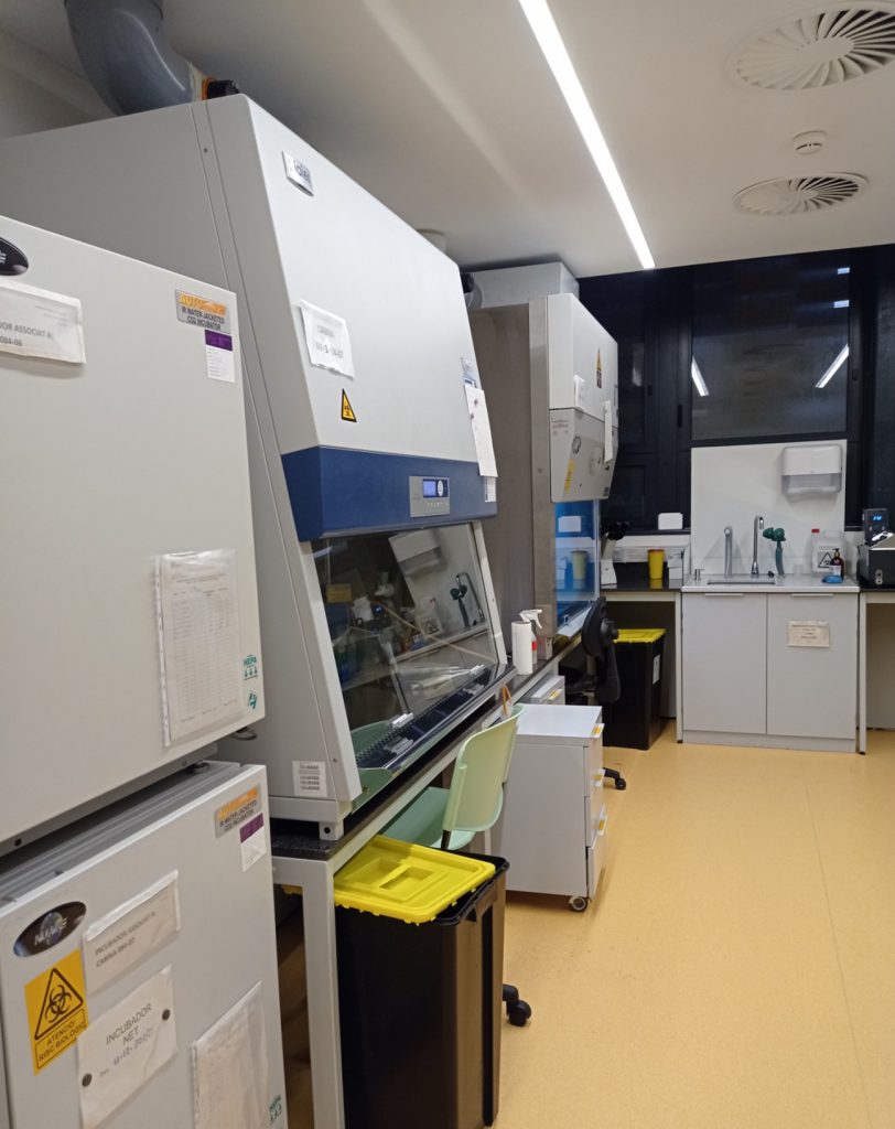
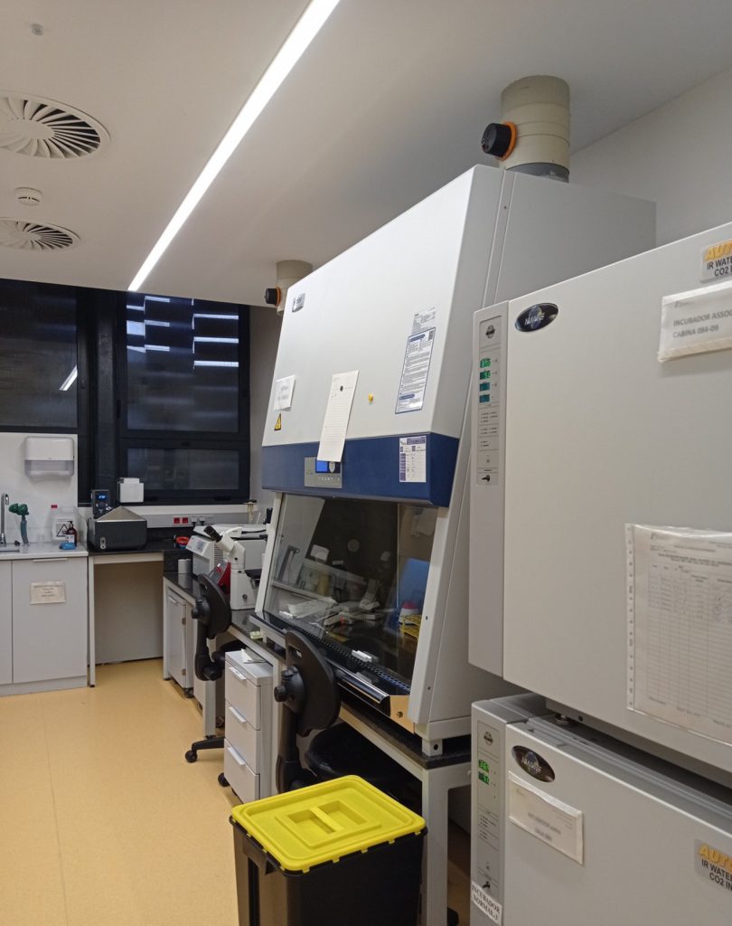
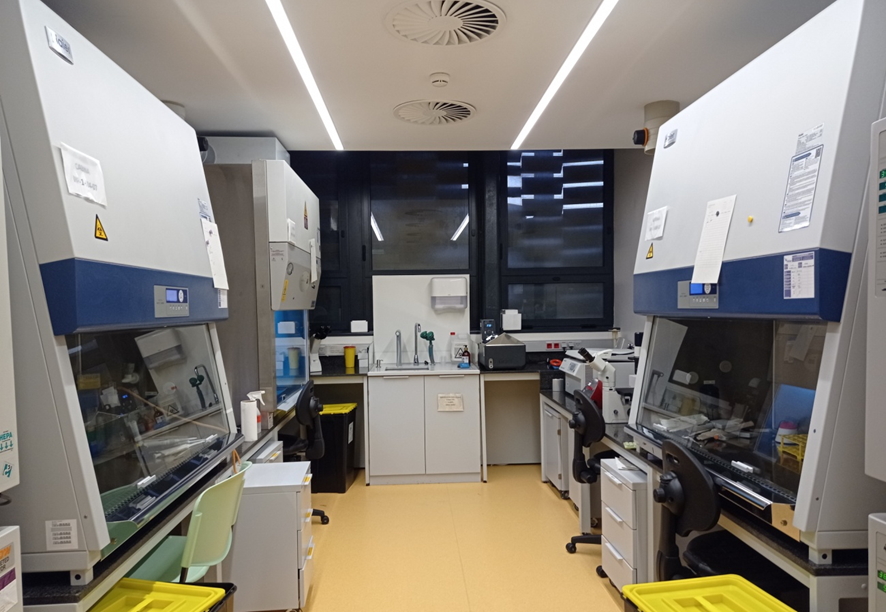
Four cell culture rooms fully with equipped with cell culture hoods, CO2 incubators, automatic cell counters, refrigerators, freezers, baths, centrifuges, inverted fluorescence and phase contrast microscopes.
Leica TCS-SP5 confocal microscope with especial features that allows studying interactions between cells/tissues and materials. Indeed, the experience of the research group in charge of this Unit makes this a unique service for the study of cells and tissues and the interactions between various materials and cell components as well as between implants/scaffolds and tissues of the recipient organism. For example, it makes it possible to characterize in detail the specific markers of particular cell populations for tissue engineering applications and in vitro tests of biocompatibility of new materials, allowing the process of cell colonization of surfaces to be explored and simultaneous analysis of the cells or subcellular structures and materials. The combined use of fluorescence and reflection makes it possible to study the tissue-implant interface, with up to 8 fluorophores, at wavelengths from 405 to 633 nm allowing histological studies of a wide range of tissue types which would be restricted at certain fixed wavelengths due to autofluorescence phenomena.
Leica TCS-SP5 confocal microscope with:
DMI 6000 inverted microscope with 4 objectives:
10X (dry); 20X; 40X and 63X(for immersion).
Confocal module:
•• 3 channels for spectral detection
•• AOBS (Acousto Optical Beam Splitter)
•• Resonant scanning system.
4 lasers:
•• Argon laser (excitation at 458, 477, 488, 496 and 514 nm)
•• He/Ne laser (excitation at 633 nm)
•• DPSS (diode-pumped solid-state) laser (excitation at 561 nm)
•• UV laser (excitation at 405 nm)
Incubation chamber on a motorized stage.
Workstation with 4 types of software for data acquisition and processing:
•• 3D imaging
•• Colocalization analysis
•• FRAP (Fluorescence Recovery After Photobleaching)
•• FLIP (Fluorescene Loss In Photobleaching)
•• FRET (Fluorescence Resonance Energy Transfer)
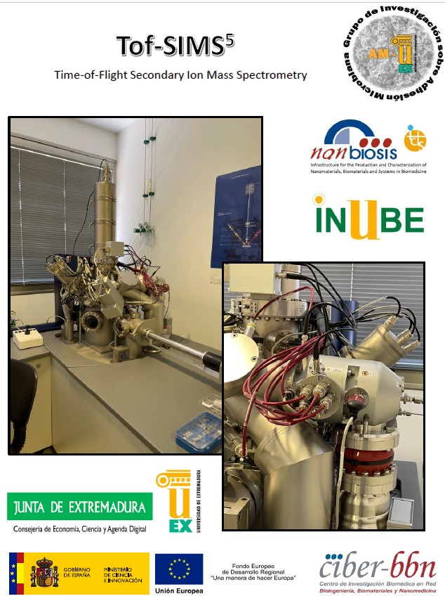
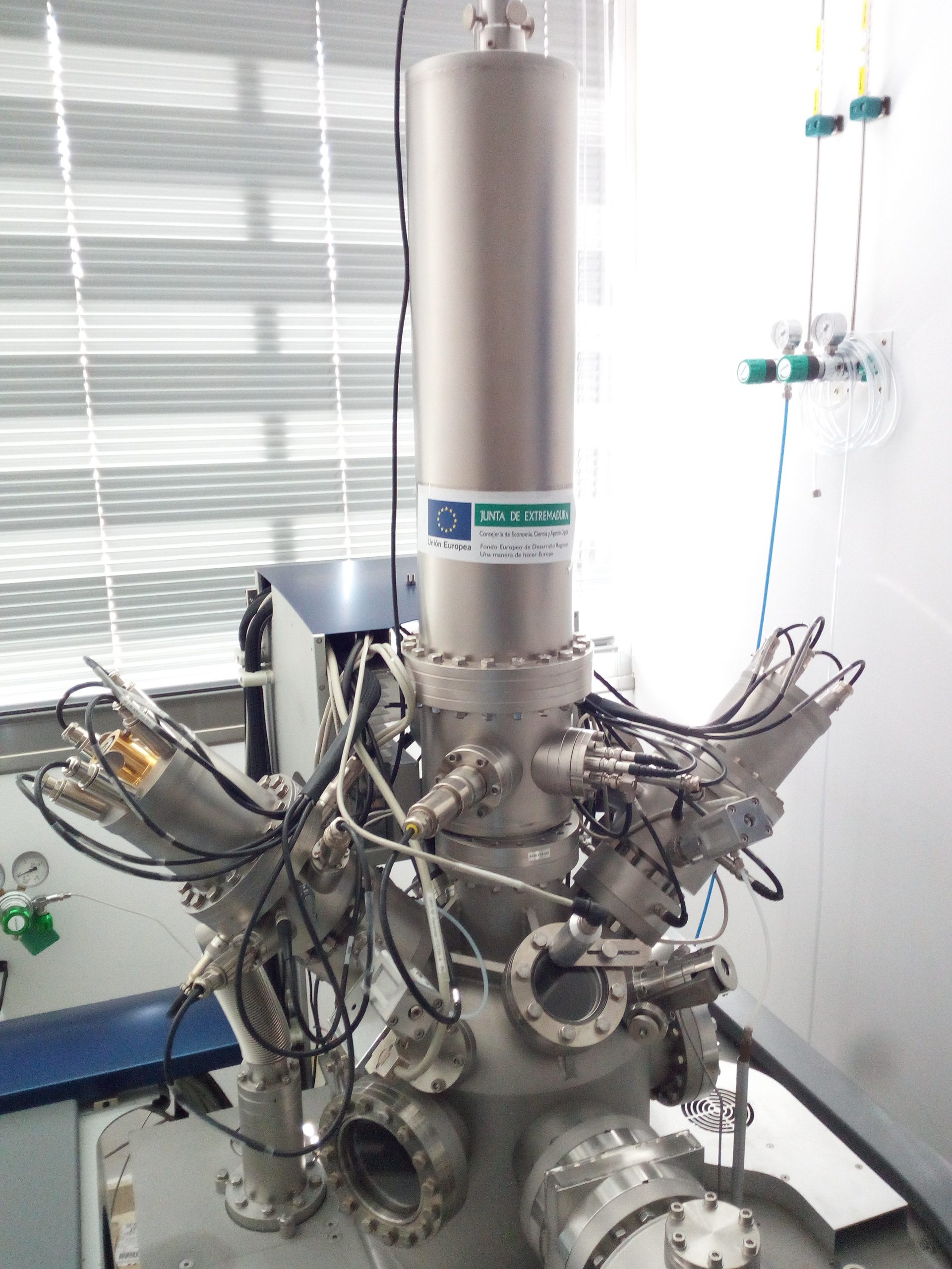
ToF-SIMS 5 Time-of-flight secondary-ion mass spectrometry system (ION TOF ).
•• Sensitivity < 100 ppm.
•• Lateral resolution ~ 100 nm (extension of surface area analyzed).
•• Depth resolution ~ 1 nm (thickness of surface analyzed).
•• Identification of molecules up to 10000 amu.
Description
X-ray photoelectron spectroscopy, known as XPS or ESCA, is one of the most widely used surface analysis techniques due to the high analytical value of its results and its flexibility in being applied to a wide variety of samples. In this regard, the surface information it provides is highly relevant, as basic XPS analysis offers qualitative and quantitative information on all the elements present on the surface of the studied material, except for hydrogen (H) and helium (He). Additionally, more comprehensive studies can provide detailed information about the surface’s chemistry, organization, and morphology.
Technical Specifications:
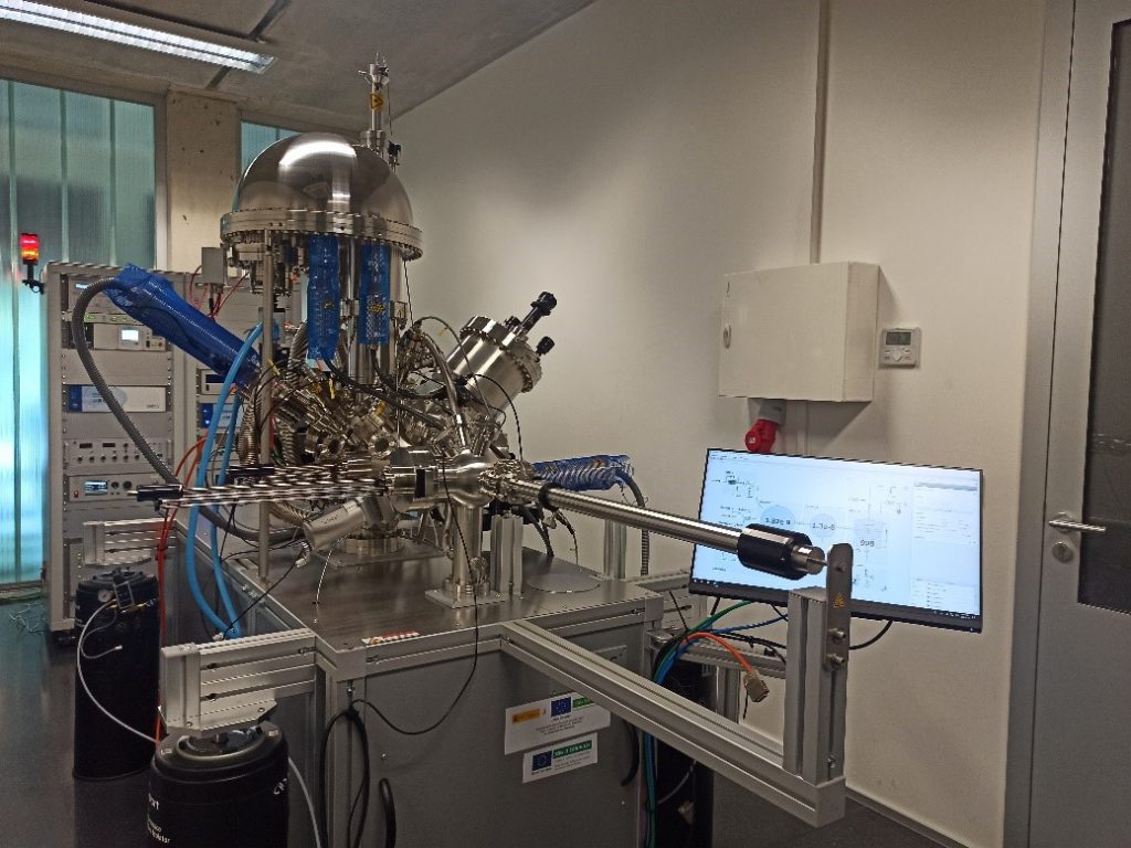
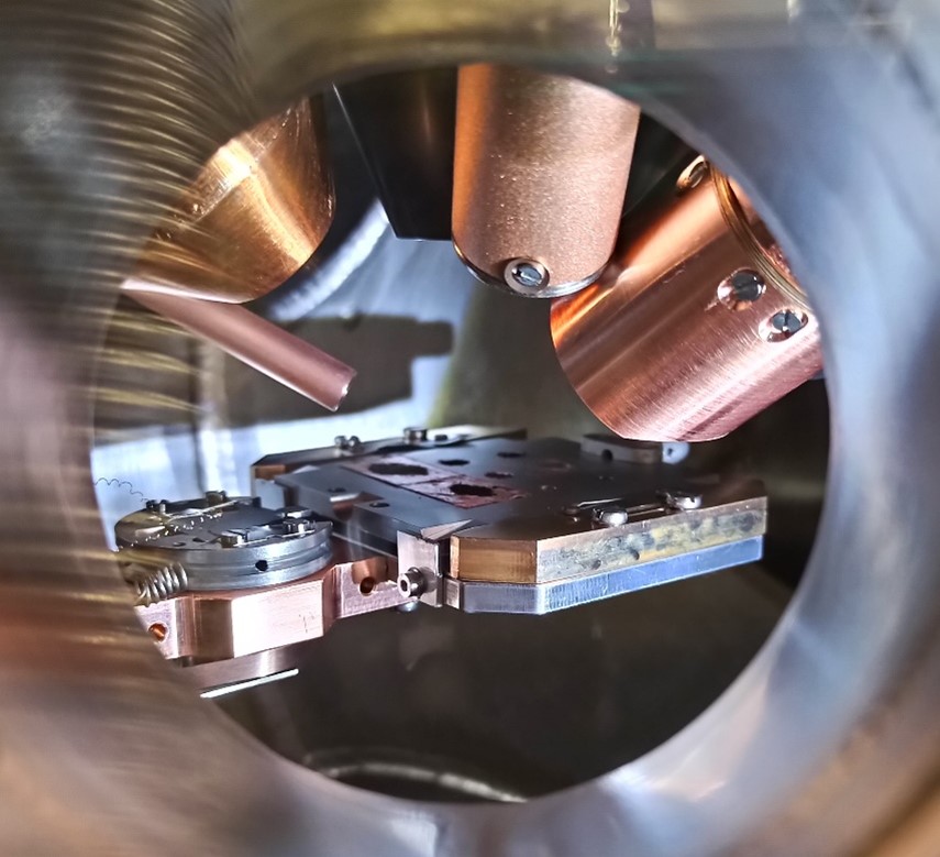
The FlexPS-ARPES-E system (SPECS) includes a UHV (ultra-high vacuum) chamber designed with a geometry that allows for the combination of the most common electronic and ionic spectroscopy applications, such as XPS, SEM/SAM (Scanning Electron/Auger scpetroscopy), UPS (Ultraviolet Photoelectron Spectroscopy), and ISS (Ion Scattering Spectroscopy). Therefore, this equipment not only allows for the collection of complete and high-resolution spectra but also includes technology for performing angle-resolved studies on layers and thin films. It also features Auger technology in SEM/SAM for acquiring elemental mappings with high lateral resolution and includes pre-chambers for thermal and gaseous treatments, enabling in situ studies of materials under their real operating conditions.
T1 and T2 Nuclear Magnetic Resonance Relaxometry:
Stelar SmarTRACER (Italy) + Bruker (Germany) 2 T electromagnet. Fast Field Cycling designed to measure longitudinal nuclear magnetic relaxation as a function of the magnetic field intensity. Measurements of the longitudinal (T1) and transverse (T2) time constants as a function of the Larmor frequency.
•• Measurement range: continuous measurement from 10 kHz (almost null field) to 10 MHz (0.25T) to obtain T1.
•• Measurement range: measurement from 10 MHz (0.25T) to 80MHz (1.9T) at desired intervals to obtain T1 and T2.
•• Inhomogeneity lower than 150 PPM.
•• Main pulse sequences implemented with the possibility of modifying parameters to the design and programming of new sequences.
•• Temperature control from -120°C to +140°C with accuracy and stability of 0.1°C.