U14-S05. Determination of cytokine concentration by flow cytometry
Determination of cytokine concentration by flow cytometry
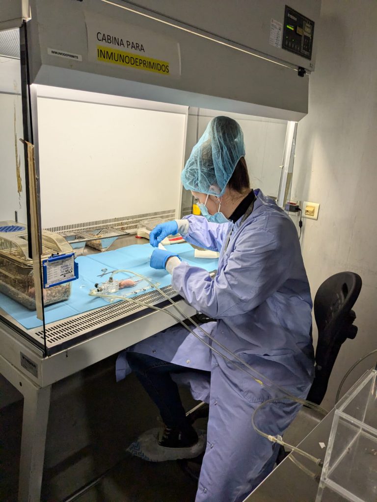
The determination of cytokine concentration by flow cytometry service represents a cutting-edge approach to cytokine analysis within the realm of biomedical research and pharmaceutical investigation. Through the utilization of advanced flow cytometry techniques and specialized staining protocols, this service enables the precise quantification of intracellular cytokines across distinct cell subsets distinguished by surface markers. This service facilitates the elucidation of cytokine profiles within specific cell populations, shedding light on their roles in health, disease, and therapeutic interventions.
Customer benefits
U14 Cell Therapy Unit offers researchers precision, efficiency, and expertise, streamlining experiment. With stringent quality control, ISO 9001-2015 and GLP compliant, tailored solutions, and access to advanced equipment, researchers can focus on innovation and core objectives, driving scientific progress forward. By delving into the intracellular milieu, researchers gain invaluable insights into the intricate regulatory networks governing immune responses and cellular signaling pathways.
Target customer
This service is essential for organizations involved in Research and Development, including pharmaceutical companies, academic institutions for biomedical research, biotechnology firms for bioprocessing, and regulatory agencies for in vitro studies.
Additional information
Selected references:
- de Pedro, Maria Ageles; Pulido, Maria; Álvarez, Veronica; Marinaro, Federica; Marchena, Ana Maria; Sanchez-Margallo, Francisco Miguel; Casado, Javier G.; Lopez, Esther. 2023. Menstrual blood-derived stromal cells: insights into their secretome in acute hypoxia conditions. MOLECULAR MEDICINE. SPRINGER. 29-1. ISSN 1076-1551. https://doi.org/10.1186/s10020-023-00646-1
- Cristobal JI; Duque, FJ; Uson-Casaus J; Barrera R; López E; Perez-Merino EM. 2022. Complete Blood Count-Derived Inflammatory Markers Changes in Dogs with Chronic Inflammatory Enteropathy Treated with Adipose-Derived Mesenchymal Stem Cells Animals. MDPI. 12-20. ISSN 2076-2615. https://doi.org/10.3390/ani12202798
- Marinaro, Federica; Silva, Joana M; Barros, Alexandre A; et al; Lopez, Esther. 2021. A Fibrin Coating Method of Polypropylene Meshes Enables the Adhesion of Menstrual Blood-Derived Mesenchymal Stromal Cells: A New Delivery Strategy for Stem Cell-Based Therapies.International journal of molecular sciences. MDPI. 22-24. ISSN 1422-0067. WOS (0) https://doi.org/10.3390/ijms222413385
- de Pedro, Maria Angeles; Gomez-Serrano, Maria; Marinaro, Federica; et al; Lopez, Esther (AC); Casado, Javier G.(4/10). 2021. IFN-Gamma and TNF-Alpha as a Priming Strategy to Enhance the Immunomodulatory Capacity of Secretomes from Menstrual Blood-Derived Stromal Cells INTERNATIONAL JOURNAL OF MOLECULAR SCIENCES. MDPI. 22-22. ISSN 1422-0067. WOS (0) https://doi.org/10.3390/ijms222212177
- López E; Blazquez R; Marinaro F; et al; Casado JG. 2019. The Intrapericardial Delivery of Extracellular Vesicles from Cardiosphere-Derived Cells Stimulates M2 Polarization during the Acute Phase of Porcine Myocardial Infarction. STEM CELL REVIEWS AND REPORTS. SPRINGER. 16-3, pp.626-626. ISSN 2629-3269. https://doi.org/10.1007/s12015-019-09926-y