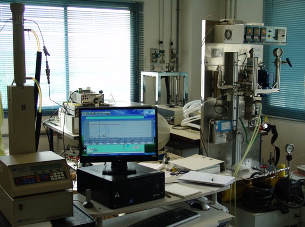
U6-E13. Ultrapyc 1200e helium pycnometer. Quantachrome
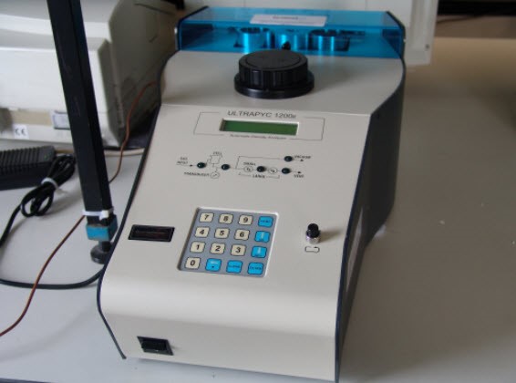 Equipment Ultrapyc 1200e Helium Pycnometer; Quantachrome Instruments
Descripción: Volume and density measurement of porous solids and powders
Especificaciones técnicas:
More info: http://www.quantachrome.com/density/auto_pycnometer.html
Aplicaciones:
Fully automated gas pycnometer, for the measurement of volume and density of powders, granular materials, and solid objects. Typical applications include: porous materials such as catalysts and activated carbons, pharmaceuticals and excipients , foods (raw, refined and end products), ceramics and refractory materials, geological samples (soils, rocks, sediments), building materials (concrete, cement), polymers and composites.