+34 620 10 75 37info@nanbiosis.com

Equipments

Equipments

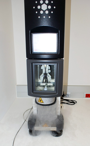
U28-E12. FEI Vitrobot Mark IV

Specifications:
Working temperature 4 – 60 °C (at an ambient temperature range between 18 – 25 °C).
Peltier controlled heating/cooling.
Maintain relative humidity at 100 %.
Ultrasonic controlled humidification.
Linux operating system.
Touch screen control and set-up. Mouse and foot pedal controls also available.
Small sample volumes can be applied manually with a pipette through a small side port on the left and the right side of the climate chamber.
Both application time and wait time (between application and blotting) are software controlled and can be set in the user interface.
Precisely timed control of multiple sample applications, blotting actions, and vitrification enables time resolved analysis of interactions among separately applied components.
Excess fluid is removed from the grid by (repeated) blotting with filter paper on rotating foam pads.
Number of blotting actions (max. 16 times for one grid) and duration of blotting are software controlled and can be set in the user interface.
Longitudinal grid positioning (‘blot offset’) and wait time between blotting and vitrification (‘drain time’) are user definable.
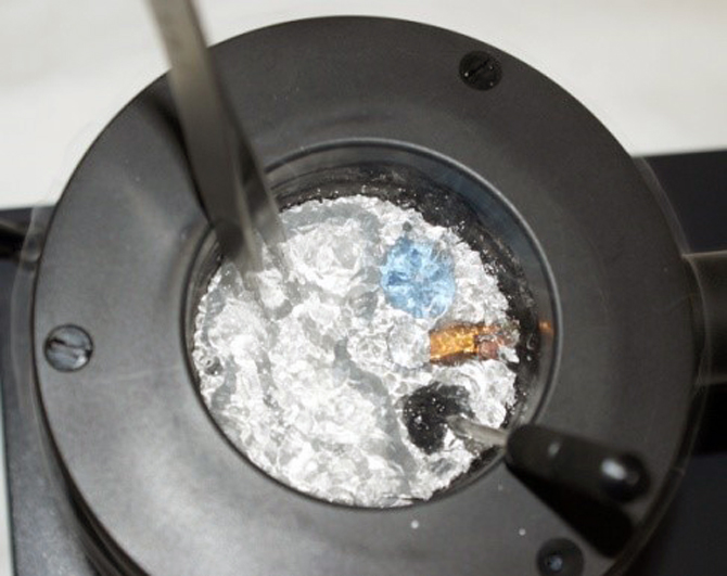
Automated shutter control allows smooth, instant injection of the sample grid into the coolant (liquid ethane or propane). A lift for the container brings the coolant close to the shutter to ensure optimal vitrification.
Synchronous lowering of the coolant container and the grid holder keeps the grid submerged in the coolant and minimizes the risk of contamination prior to transfer ring the sample into a storage box or cryo holder.
Coolant container including an integrated anti-contamination ring.

Read More

U28-E11. Leica EM AFS2

Specifications:
Reproducible operations.
Excellent specimen visibility with internal LED illumination.
Working range from -140 °C to +70 °C.
“Deep Freeze” allows sample transfer at temperatures below -140 °C.
Transfer function “TF” excludes humidity and oxygen.
Fast UV polymerization with LED UV lamp.
Any choice of substitution system, e.g.:

  • Substitution Capsule and Flat Embedding System.
  • New Microtube Embedding System EM AFS2.
  • Sapphire Disk FS System for cell cultures.
    Intuitive programming:
  • 10 users.
  • 99 programs each.
  • 99 program steps each, 0 to 99 hrs 59 mins.
  • Graphic display of substitution protocol.
  • Menu prompts.

Mouse controlled color screen.
Memory stick to transfer programs.
Log file download on memory stick.
35 liters Dewar for liquid nitrogen.
Liquid nitrogen filling from outside specimen chamber, with a consumption enough for 5 day protocols.
Stainless steel working platform.
The Leica EM FSP (“Freeze Substitution Processor”) is an automatic reagent handling system. Mounted on the Leica EM AFS2, it dispenses reagents for both FS and PLT. It automatically dilutes FS media and resins from 100 % reagent containers. An integrated LED UV lamp allows immediate polymerization of the samples.

Read More

U28-E10. Leica EM HPM100

Specifications:
• Automatic freezing of samples with one button operation.
• Extremely high cooling rates.
• Perfect freezing of samples up to 6 mm diameter.
• No cryoprotectants required.
• Rapid transfer loading device for CLEM (‘Correlative Light and Electron Microscopy’) work.
• Fast, ergonomic sample loading.
• Express sample handling for fast freezing.
• Graphical data display of actual temperature, time and pressure for each run.
• Integrated work station.
• Integrated stereomicroscope and adjustable LED illumination.
• Universal and application specific sample carriers.
• Integrated Dewar with drain outlet.
• Automatic bake-out cycle.
• Thermally insulated process chamber.
• Integrated touch control screen with prompt window.
• Freezing process cover for ultimate safety.
• USB interface for data storage and transfer.
• Integrated air compressor.

Read More

U28-E09. Leica FC7 cryo-chamber on Leica EM UC7 ultramicrotome

Specifications:
• Using the touchscreen control, three different cryo-modes are available:
– Standard.
– High gas flow, increased gaseous nitrogen flow reduces ice contamination below -140 °C.
– Wet sectioning, to set a temperature difference of up to 130 °C between knife (-40 °C) and specimen (-170 °C), which is useful for, e.g., DMSO applications.

• Leica EM CRION ionizer with electrostatic charge and discharge functions controlled by foot switches greatly improves the method of cryosection collection.
• The attachable micromanipulator provides precise positioning of the grid to make section collection easier than ever before.
• Multiple lighting angles using the patented LED chamber illumination provide excellent visibility for section manipulation and pick up.
• Contact-free, through-the-wall specimen arm makes the instrument highly stable for chatter-free cryosectioning.
• Heated chamber walls prevent icing over a prolonged working time and enhance user comfort.
• Especially for frozen hydrated sectioning or the Tokuyasu method, the Leica EM CRION with electrostatic discharge and charge mode used with the micromanipulator provides outstanding cryo sectioning performance.

Read More

U28-E08. FEI Quanta 250 FEG Scanning Electron Microscope + ESEM Mode

Specifications:
Everhart-Thornley detector of secondary electrons for high vacuum applications providing topographical information
High contrast backscattered electron detector optimized to work with low volt-ages: vCD detector. Suitable for high-vacuum and low vacuum modes, provides topographical and compositional contrast
Scanning-transmission electron microscopy detector (STEM) for high vacuum applications. Allows to introduce 8 grids in one vacuum cycle and to perform bright field and dark field analysis
Large field detector (LFD) for low vacuum and ESEM applications. Allows working in SE and BSE modes and facilitates the observation of large fields of view
Gaseous secondary electrons detector (GSED) for environmental-SEM (ESEM) applications
Gaseous analytical detector (GAD) for ESEM applications
Wet scanning-transmission electron microscopy detector (Wet-STEM) for
ESEM applications

Read More

U28-E07. FEI Tecnai G2 20 TWIN Transmission Electron Microscope + Cryo-TEM

U28-E07. FEI Tecnai G2 20 TWIN Transmission Electron Microscope + Cryo-TEM

Specifications:
Electron source with LaB6 emitter and flexible high tension between 20 and 200 kV
Twin objective lens for high contrast and high tilt applications
Side-mounted CCD Olympus Veleta with 2k x 2k MP, high field of view and fast read-out rates for real time observation
Bottom mounted CCD FEI Eagle, 4k x 4k MP, HS scintillator
EDS thanks to EDAX DPP-II analyzer with Si (Li) detector
Low-dose control software mode for electron beam damage protection of the samples.
Xplore3D software for automated tomography control.
Inspect3D software for post alignment of tomograms.
Amira software for 3D reconstruction.
Sample holders:

  • Single tilt
  • Low background (for EDS applications) double tilt
  • Fischione 2020 advanced tomography holder
  • GATAN Tomography holder for samples at room temperature.
  • GATAN Cryo-station for transferring of cryo-samples to the cryo-holder, previously vitrified of frozen with the high-pressure system.

Read More

U28-E06. Bruker Minispec MQ-60

U28-E06. Bruker Minispec MQ-60

The Minispec TD-NMR is broadly used in research, development and quality control of MRI Contrast Agents. These agents are used to enhance contrast in Magnetic Resonance images between tissues that otherwise would be difficult to differentiate. Most of these contrast agents are based on Gd chelates or iron oxide nanoparticles, which produce signal contrast enhancement by affecting the magnetic relaxation properties of water molecules. The Minispec MQ-60 provides T1 and T2 measurements with high accuracy and reproducibility, and, most importantly, at clinical field, which is fundamental for translational research. Hence, the Minispec MQ-60 plays a key role in the characterization of new contrast agents for clinical use.

Specifications:
Permanent 1.4T magnetic field.
“Intelligent” 1H probe (“plug and play”) with 60 MHz operating frequency
Fast magnet warm-up and precise digital temperature regulation for ultimate results stability
Optical detector to any kind of glass, quartz and plastic samples. Specific filters for natural and fluorescent light suppression and energy saving lamps

Read More

U28-E05. Bruker Avance 600 MHz NMR spectrometer + HR-MAS Probe

U28-E05. Bruker Avance 600 MHz NMR spectrometer + HR-MAS Probe

Specifications:
UltraShieldTM Plus 14.1 T magnet
5 mm BBFOPLUS probe, optimized for X-nuclei direct observation in a frequency range between 31P and 15N. Automated tuning and matching (ATM).
5 mm TXI probe, optimized for higher sensitivity 1H-nuclei observation. The outer coil is optimized for decoupling of 13C and 31P.Automated tuning and matching device (ATM).
TCI CryoProbe Prodigy, uses nitrogen-cooled RF coils and preamplifiers for 1H, 2H and 13C, delivering a 2.5x 1H sensitivity boost. The probe is ATM compatible and has 2H “lock” channel and Z-gradient.
Control temperature unit (from 0 ºC to 50 ºC)

Read More

U28-E04. Bruker Ascend 400 MHz NMR spectrometer

U28-E04. Bruker Ascend 400 MHz NMR spectrometer

Specifications:
UltraShieldTM Plus 9.4T magnet
5 mm BBFOPLUS probe, optimized for X-nuclei direct observation. Broad band in a frequency range between 31P and 15N. This probe has 2H “lock” channel and z gradient that allows 2D spectroscopy and hetero correlation 19F/1H and 1H/19F, to be performed. It also includes an automated tuning and matching device (ATM)
Sample Xpress Lite (for 16 samples)
Control temperature unit (from 0 ºC to 50 ºC)

Read More

U28-E03. Bruker Albira small animal CT system

U28-E03. Bruker Albira small animal CT system

Specifications:
Dedicated beds for individual rats and mice with integrated gas anesteasia.
Albira Software Suite 5.0 with integrated acquisition and tomography software.
PMOD biomedical image quantification software.
X-ray Focal Spot Size (Nominal) 35 μm.
Energy Range (kVp) 10-50.
Field of View (FOV) (transaxial x axial) 70 mm x 70mm
Multiple FOV windows can be combined for whole animal imaging
Bore Size 105 mm

Read More