+34 620 10 75 37info@nanbiosis.com

Equipments

Equipments

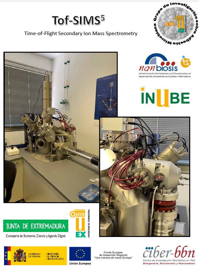
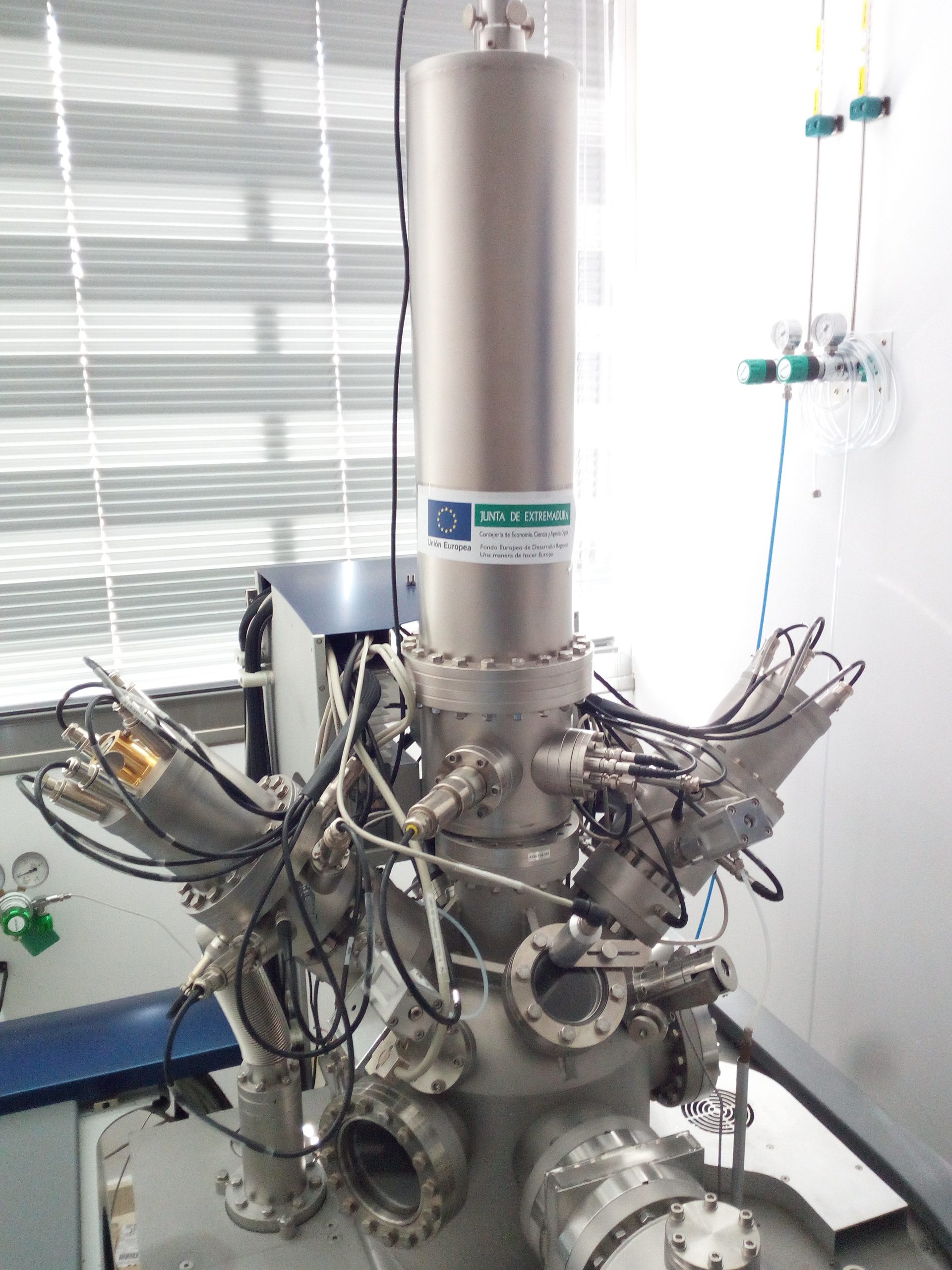
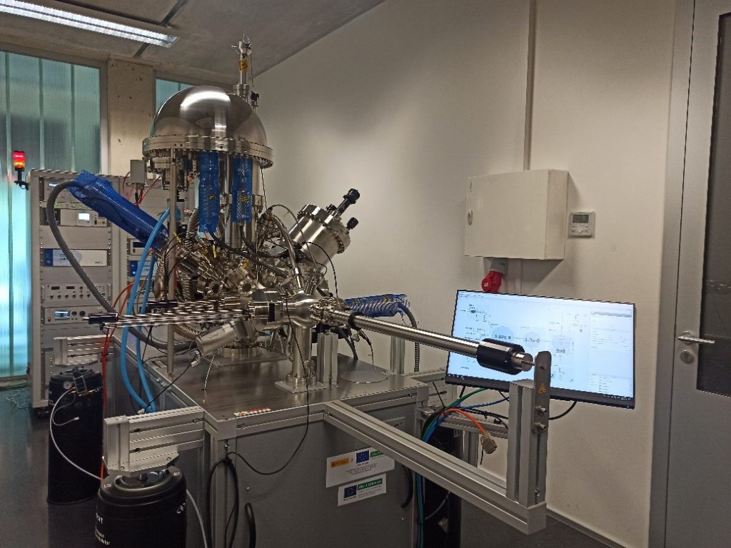
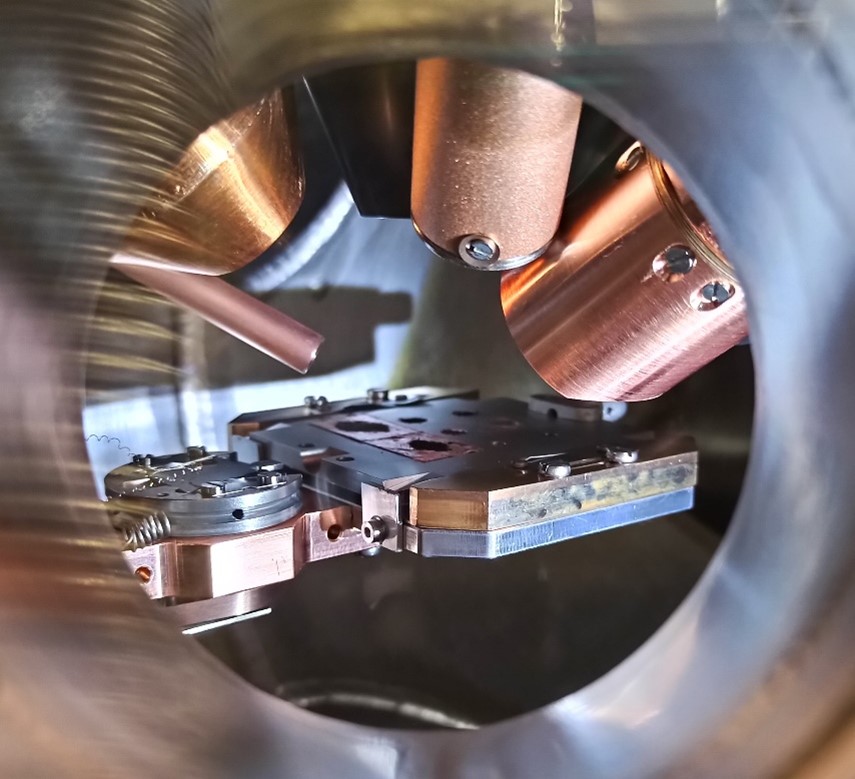
U16-E04. ToF-SIMS 5 Time-of-flight secondary-ion mass spectrometry system (ION TOF )

ToF-SIMS 5 Time-of-flight secondary-ion mass spectrometry system (ION TOF ).

•• Sensitivity < 100 ppm.
•• Lateral resolution ~ 100 nm (extension of surface area analyzed).
•• Depth resolution ~ 1 nm (thickness of surface analyzed).
•• Identification of molecules up to 10000 amu.

Read More

U16-E03. X-ray Photoelectron Spectrometer, FlexPS-ARPES-E

X-ray Photoelectron Spectrometer, FlexPS-ARPES-E

Description

X-ray photoelectron spectroscopy, known as XPS or ESCA, is one of the most widely used surface analysis techniques due to the high analytical value of its results and its flexibility in being applied to a wide variety of samples. In this regard, the surface information it provides is highly relevant, as basic XPS analysis offers qualitative and quantitative information on all the elements present on the surface of the studied material, except for hydrogen (H) and helium (He). Additionally, more comprehensive studies can provide detailed information about the surface’s chemistry, organization, and morphology.

Technical Specifications:

The FlexPS-ARPES-E system (SPECS) includes a UHV (ultra-high vacuum) chamber designed with a geometry that allows for the combination of the most common electronic and ionic spectroscopy applications, such as XPS, SEM/SAM (Scanning Electron/Auger scpetroscopy), UPS (Ultraviolet Photoelectron Spectroscopy), and ISS (Ion Scattering Spectroscopy). Therefore, this equipment not only allows for the collection of complete and high-resolution spectra but also includes technology for performing angle-resolved studies on layers and thin films. It also features Auger technology in SEM/SAM for acquiring elemental mappings with high lateral resolution and includes pre-chambers for thermal and gaseous treatments, enabling in situ studies of materials under their real operating conditions.

Read More

U15-E02. T1 and T2 Nuclear Magnetic Resonance Relaxometry

T1 and T2 Nuclear Magnetic Resonance Relaxometry:

Stelar SmarTRACER (Italy) + Bruker (Germany) 2 T electromagnet. Fast Field Cycling designed to measure longitudinal nuclear magnetic relaxation as a function of the magnetic field intensity. Measurements of the longitudinal (T1) and transverse (T2) time constants as a function of the Larmor frequency.
•• Measurement range: continuous measurement from 10 kHz (almost null field) to 10 MHz (0.25T) to obtain T1.
•• Measurement range: measurement from 10 MHz (0.25T) to 80MHz (1.9T) at desired intervals to obtain T1 and T2.
•• Inhomogeneity lower than 150 PPM.
•• Main pulse sequences implemented with the possibility of modifying parameters to the design and programming of new sequences.
•• Temperature control from -120°C to +140°C with accuracy and stability of 0.1°C.

Read More

U15-E01. Alternating Gradient Magnetometer

Alternating Gradient Magnetometer:

MicroMag M2900-4 AGM (Princeton Measurements Corporation, USA) for the magnetic and mechanical characterization of nanoparticles in various different media:
•• Magnetic Moment Range: 1nA.m² to 5mA.m² full scale (1μemu to 5emu).
•• Resolution: 0,005% of full scale with 60% overrange capability.
•• Speed of measurement: 100 ms/point.
•• High sensitivity: 10 pA.m² (10 nemu) of standard deviation at room temperature and with 1 second of averaging time.
•• High performance with samples of very small magnetization and dimensions (few nanometers).
•• The AGM can accommodate a large range of samples with widely different properties.

Read More

U5-E01. Tissue Engineering Tool 3D-300 series (nScript Inc.) for rapid prototyping

Specifications:

›› Three heads for injection of different materials:
•• Polymers
•• Ceramics
•• Living cells

›› Allowed viscosities ranging from 1 to 106 centipoises.
›› System for temperature and humidity control to enable the printing conditions to be adjusted. It is possible to create a microenvironment appropriate for each mixture of materials and proteins and to achieve high rates of cell viability.
›› Controlled by Software .
›› Visual monitoring system based on a digital camera, firewire cameras and high magnification lenses, with detection and measurement of colormetric/monochromatic variations.
›› Mapping system using a laser sensor for control of normal and extended working distances. It includes Target, Grid and Path mapping.
›› High-precision dispensing pump for dynamic control of the flow rate.

›› Position control system:
•• X/Y/Z accuracy: ±10 μm
•• X/Y reproducibility: ±2 μm
•• X/Y resolution: 0.5 μm
•• X/Y velocity: 304 mm/s (12”/s)
•• X/Y displacement: 304X 152 mm (12”X6”)
•• Z displacement: 101 mm (4”)

In addition, it should be noted that the system is sited in a clean room with a controlled environment and supply of the gases required for these tests.

Read More

U7-E07. Spin-coater

• Spin-coater

– Rotational speed: 100 to 12.000 rpm.
– Vacuum chucks: 45 and 100 mm diameters. Designed to locked substrates firmly, without deflection, and operate at very high rotational speed.
– Fragment adapters: 3 to 10 mm; 5 to 25 mm; 10 to 50 mm. Holders with an O-ring seal to maintain the vacuum level and at the same time cushion the substrate.

Read More

U7-E12. Oven

• Oven

– Temperature range: from 40°C to 250°C.
– Stability: ± 0.5°C at 100°C
– Uniformity: ± 1°C at 100°C

Read More

U7-E11. Optical Microscopy

• Optical Microscopy

– The Epi-illuminator with 12V/100w halogen lamp allows bright images to be obtained irrespective of the observation method used: brightfield, darkfield, Nomarski or polarized observations.
– Microscope objective lenses: 5x, 10x, 20x, and 100x.
– Minimum Fine focus gradation: 1 µm.
– Microscope camera head (1380×1027 pixels) ideal for image acquisition.
– Software specially designed to measure the distances of the features in the images. With these data, it is able to establish a scale bar that can be displayed on the image.

Read More

U7-E10. Profilometer

• Profilometer

– Computer controlled measurements
– High-resolution camera for color video of a 2.6 mm area.
– Variable intensity illumination for viewing samples with differing reflectivity.
– Stylus radius: 2.5 µm and 5 µm
– Stylus tracking force: 1-15 mg
– Vertical Range: up to 262 µm
– Scan Length Range: from 50 µm to 30 mm
– Vertical Resolution: down to 1 Å
– Horizontal Resolution is controlled by the scan speed
– Mechanical and optical components for sample placement, viewing and scanning.
– Manual Stage X-Y Position and sample stage rotation.

Read More

U7-E09. Interferometer

• Interferometer

– Measurement modes:
o Vertical Shift Interferometry (VSI), based on white light vertical scanning interferometry, is used for measuring features in the range of 140 nm to several µm.
o Phase Shift Interferometry (PSI), based on optical phase shifting, is dedicated to roughness measurements. Small features (1 to 140 nm) can be measured.
– The system has an automated stitching stage for large area coverage.E40
– Light Source: tungsten halogen lamp. Its brightness can be adjusted.
– 2 objective lenses included with the interferometer are available: Michelson (5.0X) and Mirau (50X).
– Field-of-View (FOV) Lenses available: 0.5, 1.0 and 2.0. FOV are lenses placed between the camera and the objective lens to adjust the field size of view.
– CCD camera to transfer the images to a computer for analysis.
– Software Vision32®. Enables advanced calculations of various surface parameters and image processing.

– Performance:
o Vertical measurement range: 0.1 nm to 1 mm
o Vertical resolution: 1 < 1 Å Ra
o Vertical scan speed up to 7.2 µm/sec
o Lateral spatial sampling 0.08 to 13.1 µm
o Field-of-View 8.24 mm to 0.05 mm (larger areas with data stitching mode)

Read More