+34 620 10 75 37info@nanbiosis.com

News U17

News U17

NANBIOSIS Strengthens Internal Collaboration at 2025 Networking Event in Cáceres

NANBIOSIS held its 2025 networking event in Cáceres to boost collaboration among Units in biomaterials, nanomedicine, and biomedical research.
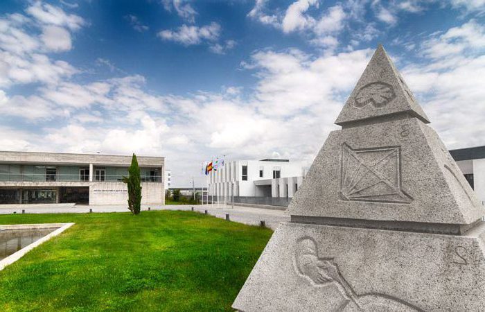
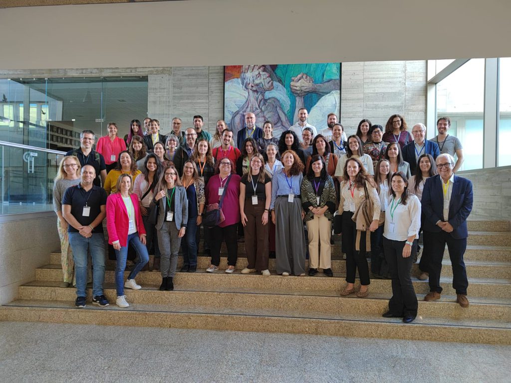
Cáceres, October 2025From 22 to 24 October 2025, the Singular Scientific and Technical Infrastructure (ICTS) NANBIOSIS held its annual internal networking event at the Centro de Cirugía de Mínima Invasión Jesús Usón (CCMIJU) in Cáceres, Spain. The event brought together Unit coordinators, researchers, and technical staff from across Spain to foster collaboration, share scientific progress, and explore new opportunities for joint projects in biomedical research, nanomedicine, and biotechnology.

A meeting to boost collaboration across NANBIOSIS Units

The three-day event was designed to enhance synergy among all the Units that make up NANBIOSIS, a distributed infrastructure offering cutting-edge services for biomaterials, nanomaterials, bioimaging, preclinical validation, and high-performance computing.

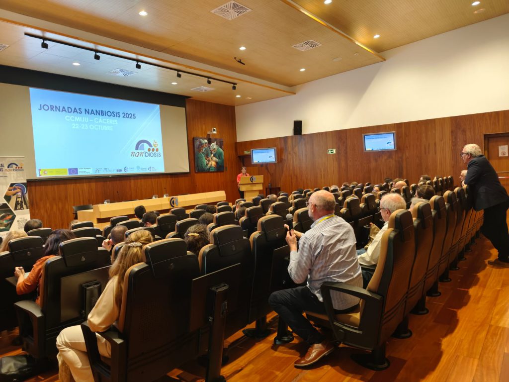
The meeting opened on 22 October with a welcome address by the Secretary General for Science of the Junta de Extremadura, who highlighted the strategic importance of research infrastructures like NANBIOSIS for innovation in health technologies. This was followed by an introduction to the initiative “Cutting Edge Biomedical Solutions”, reinforcing the mission of NANBIOSIS to support advanced biomedical development.

That afternoon, participants presented their work grouped under three scientific programmes:

  • Programme 1: Production of Biomolecules (Units 1 to 3 and 29)
  • Programme 2: Production of Biomaterials and Nanomaterials (Units 6 to 10)
  • Programme 5: High-Performance Computing (Unit 27)

The day concluded with a networking session encouraging informal exchanges and inter-Unit dialogue, in the context of the outstanding facilities of CCMIJU, one of the three nodes that form NANBIOSIS.

Guided Tour and Technical Presentations at CCMIJU

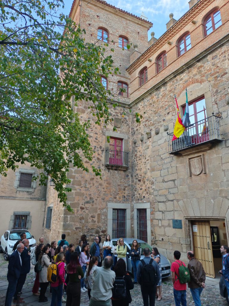
On 23 October, participants took part in a guided tour of CCMIJU, visiting the centre’s advanced research facilities in preclinical studies, imaging technologies, and tissue engineering.

After a short coffee break, the morning continued with presentations from Programme 3: Preclinical Validation – Characterisation of Tissues, Biomaterials and Surfaces (Units 12 to 19, and 30). Finally, the last programme was explained that same day in the afternoon, with Programme 4: Preclinical Validation – Bioimaging (Units 20 to 26, and 28).

Similarly to the previous day, throughout the day networking sessions facilitated direct interaction between teams, helping identify new collaboration opportunities across scientific programmes.

To close the technical sessions, participants joined “The NANBIOSIS Contest”, an interactive Kahoot-style quiz promoting engagement and friendly competition. In the evening, a guided cultural visit of Cáceres allowed attendees to explore the historic city’s UNESCO World Heritage old town.

Strengthening the NANBIOSIS Network

The final day, 24 October, saw participants returning to Madrid, concluding an event marked by enthusiasm, collaboration, and a strong sense of community across the infrastructure.

This internal networking event successfully:

  • Reinforced communication and coordination among NANBIOSIS Units.
  • Promoted awareness of each Unit’s services, equipment, and expertise.
  • Laid the groundwork for new interdisciplinary collaborations and new CEBS.
  • Highlighted the role of NANBIOSIS as a national and international reference in biomedical innovation and nanotechnology.

What is NANBIOSIS?

The goal of NANBIOSIS is to provide comprehensive and integrated advanced solutions for companies and research institutions in biomedical applications. All of this is done through a single-entry point, involving the design and production of biomaterials, nanomaterials, and their nanoconjugates. This includes their characterization from physical-chemical, functional, toxicological, and biological perspectives (preclinical validation).

Leading scientists

The main value of NANBIOSIS is our highly qualified and experienced academic scientists, working in public institutions, renowned universities and other research institutes.

Custom solutions

Designed for either scientific collaboration or the private industry, we adapt our services to your needs, filling the gaps and paving the way towards the next breakthrough.

Cutting-Edge facilities

Publicly funded, with the most advanced equipment, offering a wide variety of services from synthesis of nanoparticles and medical devices, including up to preclinical trials.

Standards of quality

Our services have standards of quality required in the pharmaceutical, biotech and medtech sectors, from Good Practices to ISO certifications.

In order to access our Cutting-Edge Biomedical Solutions with priority access, enter our Competitive Call here.

NANBIOSIS has worked with pharmaceutical companies of all sizes in the areas of drug delivery, biomaterials and regenerative medicine. Here are a few of them:

Read More

How to accomplish researchers’ goals with Confocal Microscopy: the tools, the know-how and the expertise you need

NANBIOSIS Unit 17 (Confocal Microscopy) is a CIBER-BBN unit located in the Cell Culture Unit, CAI Medicine and Biology, Faculty of Medicine at the University of Alcala. This unit of the ICTS NANBIOSIS supports researchers interested on their different studies visualizing diverse samples as tissues, cells, bacterial biofilms, etc. This unit owns the tools, the know-how and the expertise to accomplish researchers’ goals either by transmission or reflection fluorescent.

We are happy of sharing this video in which researchers of Unit 17 show all the steps required for the visualization of the PV-1 molecule, also known as PLVAP, on the gut-endothelium of cirrhotic rats. We look at the whole process, starting by the sample selection following their preparation until its visualization by the confocal fluorescent microscopy, ending up with the analyze process.

Read More

Experimental & molecular medicine: a new article with NANBIOSIS U17

ICTS-NANBIOSIS. UNIT 17 CONFOCAL MICROSCOPY SERVICE. (CIBER-BNN. UNIVERSITY OF ALCALÁ)

The UAH research groupTranslational research of chronic diseases associated with aging and kidney disease has recently published an article in which unit NANBIOSIS unit 17 and Leica SP5 confocal microscope has had a great contribution

Located in the Support Center for Research in Medicine and Biology, Faculty of Medicine and Healthe Sciences, University of Alcalá (UAH). The Unit is equipped with a Leica TCS-SP5 confocal microscope. The confocal module is equipped with three spectral detection channels, AOBS (Acousto-optical beam splitter) and a resonant scanner system that allows analysis at high speed and resolution and makes possible the analysis of dynamic in vivo physiological processes in short periods, significantly improving the survival of living biological samples by shortening the exposure times to lasers. Includes an argon laser, a He/Ne laser, a DPSS laser diode and a violet excitation laser. The microscope is coupled to a cell incubation kit that allows multi-position time-lapse experiments. The equipment includes a workstation and four software for acquisition and analysis, which allow 3D visualizations, co-location studies, FRAP (Fluorescent Recovery after Photo-bleaching), FLIP (Fluorescent Loss in Photobleaching) and FRET (Fluorescence Resonant Energy Transfer). The equipment allows 3D characterization in detail of living cells and tissues through the use of different fluorochromes, expression and localization of molecules in 2/3D, colocalization and interaction of proteins or other types of molecules; endocytosis and intracellular transport, in situ hybridization with fluorescent probes, interaction studies between cells and materials, etc.

The unit provides researchers with a wide array of routine and specialized services as well as the latest advances in microscopy, including technical and scientific support to scientists for the study of cell/tissue biology, physiology and pathogenesis of diseases.

Article of reference

Campillo, S., Bohorquez, L., Gutiérrez-Calabrés, E., García-Ayuso, D., Miguel, V., Griera, M., Calle, Y., de Frutos, S., Rodríguez-Puyol, M., Rodríguez-Puyol, D., & Calleros, L. Indoxyl sulfate- and P-cresol-induced monocyte adhesion and migration is mediated by integrin-linked kinase-dependent podosome formation Experimental & molecular medicine, 2022, 54(3): 226–238. https://doi.org/10.1038/s12276-022-00738-8

Abstract

Cardiovascular disease is an important cause of death in patients with chronic kidney disease (CKD). Protein-bound uremic toxins, such as p-cresyl and indoxyl sulfate (IS), are poorly removed during hemodialysis, leading to vascular endothelial dysfunction and leukocyte extravasation. These processes can be related to dynamic adhesion structures called podosomes. Several studies have  indicated the role of integrin-linked kinase (ILK) in the accumulation of integrin-associated proteins in podosomes. Here, we investigated the involvement of ILK and podosome formation in the adhesion and extravasation of monocytes under p-cresol (pc) and IS exposure. Incubation of THP-1 human monocyte cells with these toxins upregulated ILK kinase activity. Together, both toxins increased cell adhesion, podosome formation, extracellular matrix degradation, and migration of THP-1 cells, whereas ILK depletion with specific small interfering RNAs suppressed these processes. Interestingly, F-actin colocalized with cortactin in podosome cores, while ILK was colocalized in podosome rings under toxin stimulation. Podosome Wiskott-Aldrich syndrome protein (WASP)-interacting protein (WIP) and AKT protein depletion demonstrated that monocyte adhesion depends on podosome formation and that the ILK/AKT signaling pathway is involved in these processes. Ex vivo experiments showed that both toxins induced adhesion and podosome formation in leukocytes from wild-type mice, whereas these effects were not observed in leukocytes of conditional ILK-knockdown animals. In summary, under pc and IS stimulation, monocytes increase podosome formation and transmigratory capacity through an ILK/AKT signaling pathway-dependent mechanism, which could lead to vascular injury. Therefore, ILK could be a potential therapeutic target for the treatment of vascular damage associated with CKD.

Read More

The integrin beta1 modulator Tirofiban prevents adipogenesis and obesity by the overexpression of integrin-linked kinase: a pre-clinical approach in vitro and in vivo

Obesity is caused by the enlargement of the white adipose tissue (WAT) depots, characterized by the hypertrophic enlargement of malfunctioning adipocytes within WAT, which increases the storage of triglycerides (TG) in the lipid droplets (LD). Adipogenesis pathways as well as the expression and activity of some extracellular matrix receptors integrins are upregulated. Integrinβ1 (INTB1) is the main isoform involved in WAT remodeling during obesity and insulin resistance-related diseases. We recently described Integrin Linked Kinase (ILK), a scafold protein recruited by INTB1, as an important mediator of WAT remodeling and insulin resistance. As the few approved drugs to fight obesity have brought long-term cardiovascular side effects and given that the consideration of INTB1 and/or ILK modulation as anti-obesogenic strategies remains unexplored, we aimed to evaluate the anti-obesogenic capacity of the clinically approved anticoagulant Tirofiban (TF), stated in preclinical studies as a cardiovascular protector.

TF reduces the levels of F‑actin in adipocytes. Deprived differentiated adipocytes from c3H10T1/2 were treated with TF 50 µM or vehicle (CT) or co‑treated with specific INTB1 blocking antibody (HMB1) for the indicated times.

In the picture: Confocal images of F-actin dyed with phalloidin (red) and nuclei with DAPI (blue) of differentiated adipocytes treated with Tirofiban or vehicle (CT), where previously Integrin-beta1 was blocked with a specific blocking antibody (HMB1) for the indicated times. Scale bars 50 μm. 

F-actin confocal images were determined by the Confocal Microscopy Service of the ICTS ‘NANBIOSIS’ U17, of the Biomedical Research Networking Center on Bioengineering, Biomaterials and Nanomedicine (CIBER-BBN) with the technical assistance of Isabel Trabado, and pharmacokinetics were determined in the Applied Chemistry and Biotechnology Center (CQAB), both at Universidad de Alcalá.

Article of Reference: 

de Frutos S, Griera M, Hatem-Vaquero M, Campillo S, Gutiérrez-Calabres E, García-Ayuso D, Pardo M, Calleros L, Rodríguez-Puyol M, Rodríguez-Puyol D. The integrin beta1 modulator Tirofiban prevents adipogenesis and obesity by the overexpression of integrin-linked kinase: a pre-clinical approach in vitro and in vivo. Cell Biosci. 2022 Jan 28;12(1):10. doi: 10.1186/s13578-022-00746-1. PMID: 35090553.

Read More

“Ring Around the Rosy”

C2C12 myoblast cells differentiating into myotubes and adopting unusual shapes have been obtained and captured by resarchers of UAH and NANBIOSIS Unit 17 Confocal Microscopy Service (of CIBER-BBN and UAH) and and chosen photo of the month by the Spanish Society of Biochemistry and Molecular Biology (SEBBM)

The picture illustrates the month of October on the calendar that the SEBBM publishes and distributes annually among all partners, and is part of the gallery of images that illustrate the activities of the SEBBM. Likewise, the photography will participate in the “Best Scientific Image of the Year” contest. The winning image is awarded a grant to subsidize its attendance at the SEBBM Congress. Contributions participating in the contest will be exhibited in panels during the Congress

Authors:

Begoña Colás Escudero. Systems Biology Department. University of Alcalá. Mercedes Griera Merino. Systems Biology Department. University of Alcalá. M. Isabel Trabado Jiménez. Scientific Coordinator of NANBIOSIS – ICTS U17-Research Support Center in Medicine / Biology. UAH

Title: “Ring Around the Rosy”

The image is C2C12 myoblast cells differentiating into myotubes and adopting unusual shapes.

Blue marking: Hoescht-labeled cell nuclei.
Red marking: Cellular cytoskeleton labeled with Faloidin-Alexa 555 marking actin filaments of the cell.

For further information: here

Read More

Uraemic toxins impair skeletal muscle regeneration by inhibiting myoblast proliferation, reducing myogenic differentiation, and promoting muscular fibrosis

Elena Alcalde‑Estévez, Patricia Sosa, Ana Asenjo‑Bueno, Patricia Plaza, Gemma Olmos, Manuel Naves‑Díaz, Diego Rodríguez‑Puyol, Susana López‑Ongil & María P. Ruiz‑Torres, are the authors of an article recently published in the Journal Scientific Reports, of Nature Research, ·mentioning the collaboration in the investigation of the ICTS “NANBIOSIS” U17 Confocal Microscopy Service of CIBER-BNN and the University of Alcalá.

Uremic toxins (UT) increase in the serum in parallel with a decrease in the glomerular filtration rate and the development of sarcopenia in patients with chronic kidney disease (CKD).

This study analyses the role of UTs in sarcopenia associated with CKD in different stages of the disease.

Immunofluorescence and senescence assays were visualised using a Leica SP5 confocal microscope (Leica Microsystems, Wetzlar, Germany), through the Unit 17 Confocal Microscopy Service of the ICTS ‘NANBIOSIS’)

Through confocal microscopy studies in C2C12 cells (myoblasts), the role of high concentrations of UT in different mechanisms involved in the biology of skeletal muscle cells was observed. It was observed that they did not induce senescence (associated with beta-galactatosidase activity), but they did decrease the proliferative capacity of myoblasts, preventing the cells from entering the mitosis phase in a step prior to the condensation of chromosomes. Also, through confocal microscopy studies, it was determined that low concentrations of UT hindered myogenic differentiation of myoblasts in culture and promoted the expression of fibrosis markers” – explains Isabel Trabado, Technical Coordinator of NANBIOSIS U17

Article of reference: Alcalde-Estévez, E., Sosa, P., Asenjo-Bueno, A. et al. Uraemic toxins impair skeletal muscle regeneration by inhibiting myoblast proliferation, reducing myogenic differentiation, and promoting muscular fibrosis. Sci Rep 11512 (2021). [DOI] 

Read More

Toxicity evaluation on non-target organisms with the collaboration of NANBIOSIS U17 Confocal microscopy

Sara Jiménez-Jiménez, Georgiana Amariei, Karina Boltes, María Ángeles García and María Luisa Marina have recently published an article in the Journal of Chromatography A, ·mentioning the collaboration in the investigation of the ICTS “NANBIOSIS” U17 Confocal Microscopy Service of CIBER-BNN and the University of Alcalá.

The echocytotoxicity of racemic panthenol and dexpanthenol at different concentrations and exposure times in Spirodela polyrhiza has been studied using NANBIOSIS U17 Confocal Microscopy Service

“Panthenol (racemic mixture) and its isomer dexpanthenol have been classified as toxic for the aquatic environment by the European Regulation (EC 1272/2008). These studies are based on the natural emission (autofluorescence) of chlorophyll in different parts of the aquatic plant Spirodela polyrhiza (root, shoot and leaf). The estimation of the IC50 for each one of the compounds concludes different behavior of the compounds in the different parts of the plant, showing a higher toxicity for the racemic mixture panthenol”, explains Isabel Trabado, Technical Coordinator of NANBIOSIS U17

Article of reference:

Sara Jiménez-Jiménez, Georgiana Amariei, Karina Boltes, María Ángeles García and María Luisa Marina. Enantiomeric separation of panthenol by Capillary Electrophoresis. Analysis of commercial formulations and toxicity evaluation on non-target organisms. Journal of Chromatography A 1639 (2021) 461919. [DOI]

Financial support:

Spanish Ministry of Science and Innovation (research project PID2019-104913GB-I00). Dirección General de Universidades e Investigación de la Comunidad de Madrid (Spain), REMTAVARES (project S2018/EMT-4341). University of Alcalá for research projects CCG19/CC-068 and CCG19/IA-050, and for G.A.’s post-doctoral contract. Spanish Ministry of Science, Innovation and Universities for S.J.J.’s FPU pre-doctoral contract.

Read More

NANBIOSIS Scientific Women in the International Day of Women and Girls in Science

Today February 11 is the International Day of Women and Girls in Science, a day to raise awareness of the gender gap in science and technology.

According to the United Nations, while yet women and girls continue to be excluded from participating fully in science, science and gender equality are vital to achieve the internationally agreed development goals, including the 2030 Agenda for Sustainable Development. Thus, in recent years, the international community has made a great effort to inspire and promote the participation of women and girls in science.

NANBIOSIS wants to acknowledge  the efforts made by scientific women who struggle every day to contribute their bit to Science and highlight their essential role in nowadays research. Especially we want to recognize the work of scientists women involved in NANBIOSIS, whatever is the nature of their contribution: technical, scientific development, management, coordination, direction, etc; just to mention some examples:
Neus Ferrer and Mercedes Márquez in the Scientific Direction and Coordination of Unit 1 Protein Production Platform (PPP)
Pilar Marco and Nuria Pascual in the Management and Scientific Coordination of U2 Custom Antibody Service (CAbS) 
Miriam Royo in the Scientific Direction of U3 Synthesis of Peptides Unit
Nora Ventosa and Nathaly Segovia in the Scientific Direction and Technical Coordination of U6 Biomaterial Processing and Nanostructuring Unit
Isabel Oliveira and Teresa Galán in the Coordination of U7 Nanotecnology Unit
Rosa Villa and Gemma Gabriel in the Management and Scientific Coordination of U8 Micro – Nano Technology Unit
Gema Martínez in the Scientific Coordination of U9 Synthesis of Nanoparticles Unit
Fany Peña in the Scientific Coordination of U13 Tissue & Scaffold Characterization Unit
Mª Luisa González Martín and Margarita Hierro in the of Direction and Scientific Coordination of U16 Tissue & Scaffold Characterization Unit
Gemma Pascual and Isabel Trabado in the Coordination of the U17 Confocal Microscopy Service
Isolda Casanova in the Scientific Coordination of U18 Nanotoxicology Unit
Beatriz Moreno in the Scientific Direction of Unit 19 Clinical tests lab
Ibane Abásolo in the Scientific Coordination of Unit 20 In Vivo Experimental Platformt
Verónica Crisóstomo in the Scientific Direction of Unit 24 Medical Imaging 
Ana Paula Candiota in the Scientific Coordination of Unit 25 Biomedical Applications I 
Maria Luisa García in the Scientific Direction of U28 NanoImaging Unit from Bionand, recently incorporated to NANBIOSIS, Anna Aviñó in the Scientific Coordination of U29 Oligonucleotide Synthesis Platform (OSP) – and

Nerea Argarate in the coordination of NANBIOSIS

Thanks to all of you and your teams!

Read More

Cardiac hypertrophy research with NANBIOSIS unit 17 Confocal Microscopy Service participation

A new article has been recently published about Cardiac hypertrophy research in the Scientific Reports Jorunal by NatureResarch, counting with the expertise of NANBIOSIS unit 17 Confocal Microscopy Service form CIBER-BBN and University of Alcalá de Henares.

Cardiac hypertrophy is the abnormal enlargement, or thickening, of the heart muscle, resulting from increases in cardiomyocyte size and changes in other heart muscle components, such as extracellular matrix. This article relates more especifically with the link between long term exposure to xenoestrogen Bisphenol-A and adverse cardiovascular effects and the role of necroptosis in cardiac response to BPA had not yet been explored.

NANBIOSIS U17, Confocal microscopy Unit is mentioned in the Materials and Methods section: Slides containing tissue sections were incubated with the primary antibodies overnight 4 °C. After washing with PBS, the slides were incubated with FITC, Alexa-488, or Alexa-647-conjugated secondary antibodies for 1 hour at room temperature. Nuclei were stained with Hoechst. Images were taken for data quantification using a Leica TCS SP5 confocal microscope (UAH-NANBIOSIS-CIBER-BNN). At least five different fields per condition were obtained.

In the picture, F) Representative confocal images from heart sections of CT and 8 weeks treated BPA mice after injection with Evans blue (red fluorescence) followed by immunostaining for α-SMA (green). Nuclei were labeled with Hoechst in blue (n = 4 mice per condition). (a and b) CT hearts sections obtained at different magnifications showed no signs of EB extravasation. (c) and (d) are different sections of coronary arteries of BPA hearts with extravasation areas. Scale bar = 25 μm (a and c) and 50μm (b and d). IL = intraluminal area and (*) marks areas of EB extravasation.

Article of refrence:

Reventun, P., Sanchez-Esteban, S., Cook, A. et al. Bisphenol A induces coronary endothelial cell necroptosis by activating RIP3/CamKII dependent pathway. Sci Rep 10, 4190 (2020). https://doi.org/10.1038/s41598-020-61014-1

Read More

Optimization of Recycled-Membrane Biofilm Reactor as a sustainable biological treatment for microcystins removal with nanbiosis expertise.

Jesús Morón-López (IMDEA), Serena Molina (Chemical Engineering Department, University of Alcalá) have recently published the results of their research about Recycled-Membrane Biofilm Reactor as a sustainable biological treatment for microcystins (MC) removal. The study addresses the lack of sustainable technologies for water treatment, while opening an alternative in sustainable solid waste management under a circular economy approach.

The biofilm visualization has been performed by ICTS “NANBIOSIS”, more specifically by the Unit 17 Confocal Microscopy Service of Ciber in Bioengineering, Biomaterials & Nanomedicine (CIBER-BNN) at the Alcala University (CAI Medicine Biology) led by Juan Manuel Bellón y Gemma Pascual. In this case, the biofilm attached to the recycled membrane was observed under the confocal laser scanning microscope (CLSM Leica SP5, Leica Microsystems) of NANBIOSIS Unit 17.

Article of refrence:

Jesús Morón-López, Serena Molina, Optimization of Recycled-Membrane Biofilm Reactor (R-MBfR) as a sustainable biological treatment for microcystins removal Biochemical Engineering Journal 153 (2020) 107422

Read More