Hydrogels structured with dual stimuli responsive for biomedical applications
Researchers of NANBIOSIS Unit 17 Confocal Microscopy Service have participated in the research carried out structuring hydrophobic domains in Poly(N-isopropylacrylamide-co-Methacrylic acid) hydrogels for biomedical aplplications.
Hydrogels are cross-linked polymeric networks, which have the ability to hold a large amount of water in their structure. Hydrogels can be designed to respond to a specific stimulus such as temperature, pH, ionic strength, light, etc., making making them suitable for biomedical applications, as drug delivery.
The most popular responsive polymeric hydrogel is made of poly(N-isopropylacrylamide) (PNIPAM). The copolymerization of NIPAM with an acrylic/methacrylic acid monomer permits the development of a hydrogel with a dual stimuli response: temperature and medium pH. Additionally, the acid groups can electrostatically interact with positively charged drugs, the interaction being sensitive to pH. Therefore, these hydrogel systems have great potential for drug delivery applications.
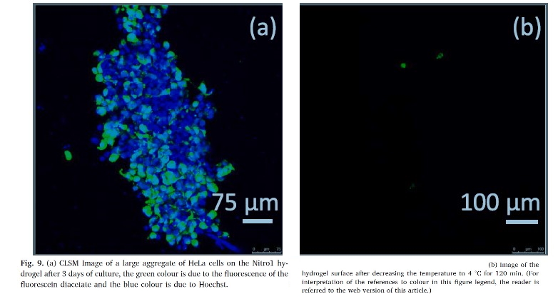
At it seemed that the structuring of dual stimuli responsive hydrogels had not been reported, the authors deat with the structuring of poly(N-isopropylacrylamide-co-methacrylic acid) hydrogels to create hydrophobic domains by means of copolymerization of NIPAM with methacrylic acid and a small percentage of a nitrocatechol monomer in an aqueous medium that contained SDS. This structured hydrogel allows is capable of loading hydrophobic molecules as well as charged drugs. The hydrogel permitted cell adhesion and growth as well as its detachment when the temperature fell below the LCST.
As reported in the article, fluorescence images of cells were obtained with a laser scanning confocal microscope (LSCM) (Leica TCS-SP5) through the Confocal Microscopy Service of ICTS ‘NANBIOSIS’ U17 of the Biomedical Research Networking Centre on Bioengineering, Biomaterials and Nanomedicine (CIBER-BBN at the University of Alcalá, Madrid, Spain).
equipped with a Diode 405 nm and a continuous Ar ion laser (488, 514,
561 and 633 nm).
Article of refrence:
Structuring hydrophobic domains in Poly(N-isopropylacrylamide-co-
Methacrylic acid) hydrogels. Mar López-González, M. Melia Rodrigo, Mercedes Valiente, Isabel Trabado, Francisco Mendicutib, Gema Marcelo. European Polymer Journal. April 2020 https://doi.org/10.1016/j.eurpolymj.2020.109695