


+34 620 10 75 37info@nanbiosis.com

Posts on Jan 1970

Iron and Magnetic Nanoparticles: Properties, Synthesis and Biomedical Applications

Introduction

Iron-based magnetic nanoparticles are among the most widely investigated nanomaterials for biomedical research. Their combination of nanoscale dimensions, magnetic responsiveness, tunable surface chemistry and potential for functionalization makes them attractive platforms for applications ranging from biomolecule delivery and regenerative medicine to biosensing, medical imaging and cancer research.

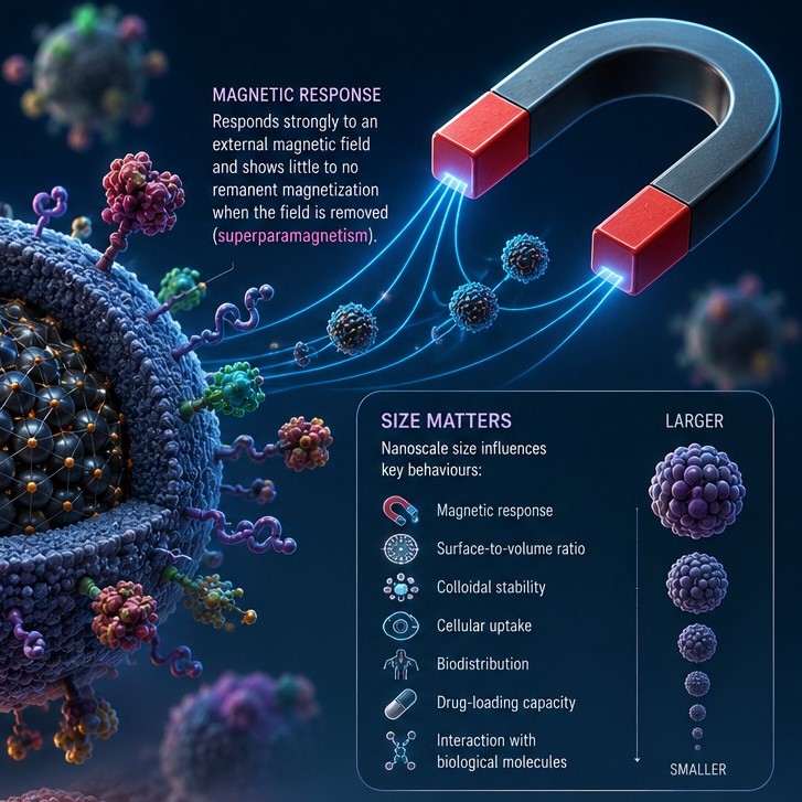
Among the different magnetic nanomaterials, iron oxide nanoparticles (IONPs)—particularly magnetite (Fe₃O₄) and maghemite (γ-Fe₂O₃)—have received considerable attention because their magnetic properties can be engineered through control of particle size, morphology, composition and surface chemistry. At sufficiently small sizes, many iron oxide nanoparticles exhibit superparamagnetic behaviour: they can respond strongly to an external magnetic field while showing little or no remanent magnetization once the field is removed. This property can help maintain colloidal stability and enables magnetic manipulation in biomedical environments.

Importantly, “magnetic nanoparticles” is a broader category than iron oxide nanoparticles. Magnetic nanoparticles can contain different magnetic materials, including ferrites and other compositions. However, iron oxide nanoparticles are particularly relevant to biomedicine because of their combination of magnetic properties, established synthesis methods and the possibility of surface functionalization.

Research into these materials is advancing toward increasingly sophisticated multifunctional and theranostic systems, in which a single nanosystem can combine diagnostic, targeting and therapeutic functions.

What are Magnetic Nanoparticles?

Magnetic nanoparticles (MNPs) are nanoparticles that respond to an external magnetic field. Their magnetic behaviour depends strongly on their composition, crystal structure, size and morphology.

In biomedical research, one of the most important groups is magnetic iron oxide nanoparticles, including:

  • Magnetite (Fe₃O₄)
  • Maghemite (γ-Fe₂O₃)
  • Other iron-containing magnetic formulations and composite systems

At the nanoscale, iron oxide particles can display superparamagnetism, a property that is particularly useful for biomedical applications. Superparamagnetic nanoparticles can be attracted or manipulated using an external magnetic field but, after the field is removed, do not necessarily retain a permanent magnetic moment. This reduces magnetic aggregation compared with permanently magnetized particles under appropriate conditions.

Why does nanoparticle size matter?

The physical behaviour of magnetic nanoparticles is strongly influenced by their size. Changes in particle size can affect:

  • Magnetic response
  • Surface-to-volume ratio
  • Colloidal stability
  • Cellular uptake
  • Biodistribution
  • Drug-loading capacity
  • Interaction with biological molecules

Particle morphology and surface chemistry are also important. For example, nanoparticles can be coated or functionalized with polymers, silica, lipids, proteins or other molecules to improve colloidal stability, reduce unwanted interactions and introduce specific biological functions.

This surface functionalization is particularly important when magnetic nanoparticles are being developed as nanocarriers, because therapeutic molecules, targeting ligands or other biomolecules can be incorporated into or attached to the nanoparticle system.

Advantages of Magnetic Nanoparticles

Magnetic nanoparticles offer several characteristics that make them attractive for biomedical applications.

Magnetic manipulation

One of their defining advantages is the possibility of manipulating nanoparticles using an external magnetic field. Depending on the application and experimental configuration, magnetic fields can be used to concentrate, separate, guide or retain magnetic nanoparticles. This principle is particularly interesting for targeted delivery and cell separation.

Surface functionalization

The surface of iron oxide nanoparticles can be chemically modified to attach or encapsulate different molecules. This provides considerable flexibility for designing nanosystems for specific biological applications.

Surface modification can also influence:

  • Stability in biological fluids
  • Protein adsorption
  • Cellular interactions
  • Biocompatibility
  • Drug loading
  • Targeting behaviour

Consequently, the same magnetic core can be incorporated into very different nanoplatforms depending on the intended application.

Multifunctionality

Magnetic nanoparticles can combine several functions within one platform. For example, a nanoparticle can potentially act as a carrier for a therapeutic molecule while also providing a magnetic or imaging function.

This combination of diagnosis and therapy is one of the foundations of nanotheranostics.

Tunable physicochemical properties

Size, morphology, composition, coating and surface chemistry can be adjusted during nanoparticle synthesis and post-synthesis functionalization. These parameters influence the magnetic, colloidal and biological properties of the final material.

Potential for biomedical integration

Iron oxide nanoparticles have been investigated in drug delivery, magnetic resonance imaging, biosensing, cell labelling, tissue engineering and cancer research. Nevertheless, their suitability for a specific biomedical application must always be evaluated experimentally, since nanoparticle behaviour depends strongly on formulation, dose, surface chemistry and biological environment.

How are Magnetic Nanoparticles Synthesized?

The synthesis method has a major influence on the final characteristics of magnetic nanoparticles. Researchers seek to control parameters such as particle size, size distribution, morphology, crystallinity, magnetic properties, surface chemistry and colloidal stability.

Several approaches are available.

Co-precipitation

Co-precipitation is one of the most commonly used approaches for preparing iron oxide nanoparticles. It generally involves the precipitation of iron ions under controlled chemical conditions, producing iron oxide nanoparticles in an aqueous or aqueous-compatible environment.

The method is relatively straightforward and can be suitable for producing nanoparticles at scale. However, controlling particle size distribution, morphology and oxidation state can be challenging.

Thermal decomposition

Thermal decomposition involves the decomposition of suitable iron-containing precursors at elevated temperatures, often in the presence of stabilizing ligands or surfactants.

Compared with conventional co-precipitation, thermal decomposition can provide greater control over particle size, morphology and crystallinity. However, it may require organic solvents, higher temperatures and additional processing steps for biomedical applications.

Hydrothermal and solvothermal synthesis

Hydrothermal and solvothermal approaches use controlled temperature and pressure conditions to produce nanoparticles with specific structural and morphological characteristics. These methods can be useful when precise control of particle properties is required.

Sol-gel, microemulsion and other approaches

Other synthesis strategies include:

  • Sol-gel methods
  • Microemulsion techniques
  • Sonochemical synthesis
  • Microwave-assisted synthesis
  • Biological or biomimetic approaches

Each technique offers different advantages and limitations in terms of scalability, reproducibility, size control, morphology and surface chemistry.

Surface functionalization

Synthesis is only one part of developing a biomedical magnetic nanoparticle. The particles are frequently coated or functionalized after synthesis.

Possible surface modifications include polymers, silica, lipids, ligands and biomolecules. Functionalization can improve colloidal stability and biocompatibility and can introduce specific interactions with cells or biological targets.

For biomedical applications, characterization of the final functionalized nanoparticle—not just the magnetic core—is therefore essential.

Biomedical Applications of Magnetic Nanoparticles

Magnetic nanoparticles are being investigated across a wide range of biomedical applications. Some are already well established as research tools, while others remain primarily at the preclinical or experimental stage.

1. Biomolecule Delivery and Gene Therapy

One of the most promising applications of magnetic nanoparticles is the delivery of biomolecules, including nucleic acids and therapeutic molecules.

Magnetic nanoparticles can be incorporated into nanocarriers that transport a biological payload. An external magnetic field can potentially influence the spatial distribution of magnetic carriers, creating opportunities for magnetically assisted targeting and retention.

Gene delivery

Magnetic nanoparticles have also been investigated as non-viral vectors for gene delivery. Nucleic acids such as DNA, RNA or other gene-regulating molecules can be associated with appropriately functionalized nanoparticle systems.

The objective is to protect the nucleic acid, facilitate its transport and promote delivery to target cells. Magnetic guidance can be incorporated into these systems as an additional targeting strategy.

Research in this area includes delivery systems for:

  • DNA
  • siRNA
  • mRNA
  • Other therapeutic nucleic acids

However, successful gene delivery requires much more than magnetic targeting. Cellular uptake, endosomal escape, intracellular trafficking, payload release and potential toxicity all need to be addressed when designing a magnetic gene-delivery system.

Controlled drug and biomolecule release

Magnetic nanoparticles can also be integrated into systems designed for controlled release. Depending on the formulation, release can be influenced by environmental conditions or external stimuli, including magnetic fields.

This creates opportunities for developing multifunctional nanocarriers in which targeting, delivery and imaging are combined.

For researchers developing new delivery systems, the magnetic core can therefore serve as one component of a larger engineered nanoplatform rather than functioning as the therapeutic agent itself.

2. Regenerative Medicine

Magnetic nanoparticles are increasingly investigated in tissue engineering and regenerative medicine.

Their magnetic properties allow researchers to manipulate cells or biomaterials using external magnetic fields. For example, magnetic nanoparticles can be incorporated into biomaterial systems or associated with cells to influence their positioning, organization or interaction with engineered scaffolds.

Research has explored magnetic approaches for:

  • Cell manipulation and positioning
  • Tissue engineering
  • Stem-cell research
  • Cell patterning
  • Tissue repair
  • Magnetic stimulation of cellular environments

Magnetic nanoparticles can also be incorporated into composite biomaterials to provide additional functionality while maintaining the structural role of the scaffold.

The field is particularly interesting because magnetic control can introduce a degree of remote manipulation that conventional biomaterials do not provide.

3. Magnetic Sensors and Biosensors

Magnetic nanoparticles can be used in sensing and biosensing technologies because biological recognition events can be coupled to changes in magnetic properties or to magnetic separation and concentration.

Functionalized nanoparticles can bind specific biomolecules, cells or pathogens. Their magnetic properties can then facilitate separation, concentration or detection.

Applications investigated include the detection of:

  • Proteins
  • Nucleic acids
  • Biomarkers
  • Cells
  • Pathogens
  • Small molecules

Iron oxide nanoparticles have also been investigated in more complex biosensor platforms, including systems combining magnetic nanoparticles with optical or electronic detection mechanisms.

4. Diagnostic Imaging

Magnetic iron oxide nanoparticles have been extensively investigated for medical imaging, particularly magnetic resonance imaging (MRI).

Their magnetic properties can influence the relaxation behaviour of nearby water protons, enabling their use as contrast agents in appropriate formulations. The imaging behaviour depends on particle size, magnetic properties, surface characteristics and the specific MRI sequence being used.

Iron oxide nanoparticles have also been investigated for other imaging approaches, including magnetic particle imaging (MPI). However, it is important to distinguish promising research applications from technologies that are routinely used clinically: the clinical translation and regulatory status of specific nanoparticle formulations vary considerably.

5. Nanomedicine and Theranostics

The multifunctionality of magnetic nanoparticles makes them attractive platforms for nanomedicine.

A single nanosystem can potentially combine:

  • Targeting
  • Drug delivery
  • Imaging
  • Magnetic manipulation
  • Stimulus-responsive release
  • Therapeutic activity

This creates the possibility of theranostic platforms, designed to combine diagnostic and therapeutic functions.

For example, a magnetic nanoparticle system could be engineered to carry a therapeutic molecule while simultaneously providing an imaging signal. Such approaches are being extensively investigated, particularly in oncology.

6. Catalysis in the Tumour Environment

Iron oxide nanoparticles are also being investigated as catalytic nanomaterials in the context of cancer research.

Some iron-containing nanoparticles can participate in reactions involving reactive oxygen species. This property is being explored in nanocatalytic cancer therapies, including approaches related to chemodynamic therapy.

A major research objective is to exploit characteristics of the tumour microenvironment—such as altered pH or the presence of endogenous hydrogen peroxide—to promote localized chemical reactions capable of generating cytotoxic species.

However, these approaches remain an active area of research, and their efficacy and safety depend strongly on nanoparticle composition, localization and the biological environment.

7. Artificial Enzymes and Nanozymes

Some iron oxide nanoparticles exhibit enzyme-mimetic catalytic activity. Nanomaterials with enzyme-like properties are commonly known as nanozymes.

Iron oxide nanoparticles have been reported to exhibit activities resembling those of natural peroxidase enzymes under appropriate conditions. This has generated interest in applications such as biosensing, diagnostics and catalytic biomedical systems.

The catalytic behaviour of a nanozyme depends on several factors, including particle size, surface chemistry, composition and the surrounding chemical environment.

This field illustrates how magnetic nanoparticles can provide more than magnetic functionality: the same nanomaterial can potentially combine magnetic manipulation with catalytic activity, opening opportunities for multifunctional biomedical platforms.

Frequently Asked Questions

What are magnetic nanoparticles?

Magnetic nanoparticles are nanoscale materials that respond to an external magnetic field. In biomedicine, iron oxide nanoparticles such as magnetite and maghemite are among the most extensively studied examples.

What are iron oxide nanoparticles used for?

Iron oxide nanoparticles are investigated for drug and biomolecule delivery, gene delivery, regenerative medicine, biosensing, medical imaging, cell separation, cancer research, magnetic hyperthermia and nanozyme-based applications.

What is superparamagnetism?

Superparamagnetism is a nanoscale magnetic behaviour in which sufficiently small magnetic particles can respond strongly to an external magnetic field but show little or no permanent magnetization after the field is removed. It is an important property for many biomedical magnetic nanoparticle systems.

Can magnetic nanoparticles deliver drugs?

Yes. Magnetic nanoparticles can be incorporated into drug-delivery systems and can potentially be guided or concentrated using external magnetic fields. Their surfaces can also be functionalized to improve drug loading, stability and targeting.

Can magnetic nanoparticles be used for gene delivery?

Yes. Functionalized magnetic nanoparticles have been investigated as non-viral carriers for DNA, RNA and other nucleic acids. Research focuses on improving cellular uptake, intracellular trafficking, release and biological safety.

Are magnetic nanoparticles safe?

Safety depends on the specific nanoparticle formulation, including its composition, size, surface coating, dose, route of administration and biological fate. Although iron oxide nanoparticles have important biomedical advantages, questions regarding biodistribution, metabolism, protein adsorption and toxicity must be evaluated for each formulation and application.

How are magnetic nanoparticles synthesized?

Common synthesis approaches include co-precipitation, thermal decomposition, hydrothermal or solvothermal synthesis, sol-gel methods, microemulsion and sonochemical techniques. The choice of method influences particle size, morphology, crystallinity and magnetic properties.

What is a nanozyme?

A nanozyme is a nanomaterial with enzyme-like catalytic activity. Iron oxide nanoparticles are among the materials investigated as nanozymes, including for biosensing and catalytic biomedical applications.

Conclusion

Magnetic iron oxide nanoparticles combine nanoscale properties with magnetic responsiveness and a highly engineerable surface, making them versatile materials for biomedical research.

Their potential extends from biomolecule and gene delivery to regenerative medicine, biosensing, diagnostic imaging, nanomedicine, tumour-associated catalysis and nanozyme applications. Particularly attractive is their ability to integrate several functions into a single platform, enabling researchers to explore systems that combine targeting, delivery, imaging and therapy.

At the same time, the development of biomedical magnetic nanoparticles requires careful control of particle size, morphology, magnetic properties, surface chemistry, colloidal stability and biological interactions. Promising laboratory results do not automatically translate into clinical applications, and safety, pharmacokinetics, reproducibility, scalability and regulatory requirements remain important considerations.

For researchers and companies developing advanced nanomedicine systems, the ability to design, synthesize, functionalize and comprehensively characterize magnetic nanoparticles is therefore essential. This multidisciplinary approach is key to translating magnetic nanomaterials from experimental platforms into reliable biomedical technologies.

Related Articles

What is NANBIOSIS?

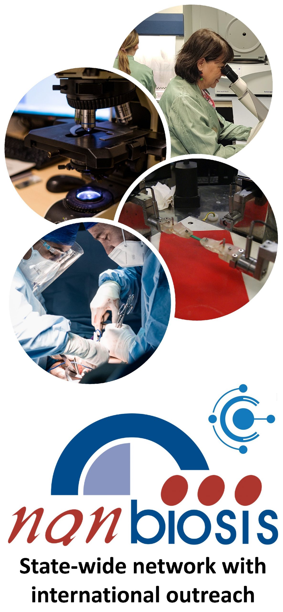
The goal of NANBIOSIS is to provide comprehensive and integrated advanced solutions for companies and research institutions in biomedical applications. All of this is done through a single-entry point, involving the design and production of biomaterials, nanomaterials, and their nanoconjugates. This includes their characterization from physical-chemical, functional, toxicological, and biological perspectives (preclinical validation).
Leading scientists

The main value of NANBIOSIS is our highly qualified and experienced academic scientists, working in public institutions, renowned universities and other research institutes.

Custom solutions

Designed for either scientific collaboration or the private industry, we adapt our services to your needs, filling the gaps and paving the way towards the next breakthrough.

Cutting-Edge facilities

Publicly funded, with the most advanced equipment, offering a wide variety of services from synthesis of nanoparticles and medical devices, including up to preclinical trials.

Standards of quality

Our services have standards of quality required in the pharmaceutical, biotech and medtech sectors, from Good Practices to ISO certifications.

In order to access our Cutting-Edge Biomedical Solutions with priority access, enter our Competitive Call here.

NANBIOSIS has worked with pharmaceutical companies of all sizes in the areas of drug delivery, biomaterials and regenerative medicine. Here are a few of them:

Read More

Gold Nanoparticles: Properties, Synthesis and Biomedical Applications

Introduction

Gold nanoparticles (AuNPs) are among the most extensively studied inorganic nanomaterials in nanomedicine and biomedical research. Their combination of tunable size and shape, distinctive optical properties, high surface-to-volume ratio and versatile surface chemistry makes them valuable platforms for designing systems for drug and biomolecule delivery, biosensing, imaging and therapeutic applications.

Unlike bulk gold, gold at the nanoscale exhibits physicochemical properties that can be strongly influenced by particle size, morphology, surface chemistry and the surrounding environment. In particular, gold nanoparticles display localized surface plasmon resonance (LSPR), an optical phenomenon that gives rise to strong and size- and shape-dependent interactions with light. This property can be exploited for sensing, imaging and photothermal applications.

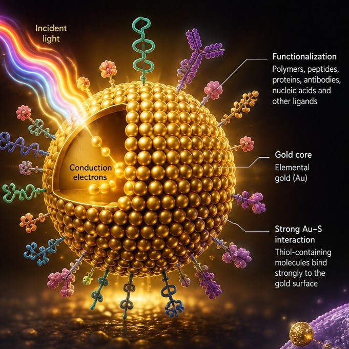
Another important feature is the ability to functionalize the gold surface with a wide variety of molecules, including polymers, peptides, proteins, antibodies and nucleic acids. This makes AuNPs particularly attractive as multifunctional platforms that can combine delivery, targeting, sensing and therapeutic functions within the same nanosystem.

For these reasons, gold nanoparticles continue to attract considerable interest in nanomedicine, drug delivery, gene therapy, regenerative medicine, biosensing and cancer research. However, many proposed biomedical applications remain at the experimental or preclinical stage, and factors such as biodistribution, clearance, long-term safety, surface chemistry and manufacturing reproducibility remain important considerations for clinical translation.

What are Gold Nanoparticles?

Gold nanoparticles are nanoscale structures composed primarily of elemental gold (Au). They can be produced in different sizes and morphologies, including spherical nanoparticles, nanorods, nanoshells, nanostars, nanocages and other anisotropic structures.

Their properties depend strongly on their physical and chemical characteristics. Important parameters include:

  • Particle size: influences cellular uptake, biodistribution, optical properties and interactions with biological systems.
  • Shape and morphology: determine optical and plasmonic behaviour and can influence biological interactions.
  • Surface chemistry: affects colloidal stability, protein adsorption, cellular interactions and biological fate.
  • Surface charge: can influence interactions with cell membranes and biomolecules.
  • Ligand or coating: polymers, peptides, antibodies, nucleic acids and other molecules can be attached to the gold surface to provide targeting or functional properties.

One of the most characteristic properties of AuNPs is their localized surface plasmon resonance. At the appropriate nanoscale dimensions, incident light can interact with the conduction electrons of the gold nanoparticle, producing strong optical absorption and scattering. The resulting optical response changes with particle size, shape, aggregation state and surrounding medium.

The gold surface can also interact strongly with thiol-containing molecules, providing a widely used route for surface functionalization. Together with other conjugation strategies, this enables the construction of AuNP-based systems carrying therapeutic molecules, targeting ligands or recognition elements.

Advantages of Gold Nanoparticles

The interest in gold nanoparticles for biomedical applications comes from several complementary advantages.

Tunable physicochemical properties

AuNPs can be engineered with different sizes, shapes and surface chemistries. These parameters can be adjusted to modify their optical, colloidal and biological behaviour.

Unique optical properties

Their LSPR gives AuNPs distinctive optical responses. By modifying particle size and morphology, researchers can tune absorption and scattering properties, including responses in regions of the spectrum relevant to biological applications. Gold nanostructures designed to interact with near-infrared light are particularly interesting for photothermal approaches because near-infrared wavelengths can penetrate biological tissues more effectively than visible light.

High surface area and versatile functionalization

The large surface area relative to their volume allows numerous molecules to be associated with an individual nanoparticle. AuNPs can therefore be functionalized with drugs, nucleic acids, peptides, proteins, antibodies, polymers and other ligands.

Multifunctionality

A single AuNP platform can potentially combine several functions, such as biomolecule delivery, molecular recognition, imaging and photothermal activity. This multifunctionality is one of the reasons AuNPs are being investigated for theranostic applications, in which diagnosis and therapy are integrated into a single platform.

Catalytic and enzyme-mimicking properties

Gold nanoparticles can exhibit catalytic activity and, depending on their size, morphology and surface chemistry, can show enzyme-like activities. These materials are often referred to as gold nanozymes. Reported enzyme-mimicking activities include peroxidase-, oxidase-, catalase- and superoxide-dismutase-like behaviour.

At the same time, these advantages should not be interpreted as evidence that all AuNP formulations are inherently safe or clinically suitable. Their biological effects depend substantially on parameters such as size, shape, surface coating, dose, aggregation and exposure route. Understanding these structure–activity relationships is therefore essential for biomedical development.

How are Gold Nanoparticles Synthesized?

Gold nanoparticles can be synthesized using several chemical, physical and biological approaches. The selected method depends on the desired particle size, morphology, surface chemistry, dispersity and intended application.

Chemical synthesis

A widely used approach is the Turkevich method, in which a gold precursor such as chloroauric acid (HAuCl₄) is reduced in the presence of citrate. The process involves the formation of gold nuclei followed by particle growth. Reaction conditions influence nucleation and growth and therefore affect the resulting particle size and distribution.

Other chemical approaches include:

  • Seed-mediated growth, which allows controlled growth of nanoparticles from preformed seeds and is particularly useful for producing anisotropic structures such as nanorods.
  • Sodium borohydride reduction, which can produce small gold nanoparticles under appropriate reaction conditions.
  • Brust–Schiffrin synthesis, commonly used to prepare thiol-protected gold nanoparticles.
  • Ascorbic-acid-based reduction methods, used in several controlled-growth approaches.

Biological or “green” synthesis

Biological synthesis uses biological materials or extracts as reducing and/or stabilizing agents. Plant extracts, microorganisms and biomolecules have all been investigated for the production of gold nanoparticles.

These approaches can reduce the use of some conventional chemical reagents, but biological synthesis can also introduce challenges related to batch-to-batch variability, composition and reproducibility. Consequently, the most appropriate synthesis route depends on the requirements of the final biomedical application.

Surface functionalization

Synthesis is only one part of AuNP engineering. For biomedical applications, the nanoparticle surface is frequently modified after synthesis or functionalized during preparation.

Surface coatings can improve colloidal stability and provide functional groups for attaching biological molecules. For example, AuNPs can be functionalized with nucleic acids, peptides, proteins, antibodies or polymers. Such modifications can influence cellular uptake, biodistribution, targeting and the interaction of the nanoparticles with biological fluids.

Biomedical Applications of Gold Nanoparticles

1. Biomolecule Delivery and Gene Therapy

One of the most important research applications of gold nanoparticles is the delivery of biomolecules, including nucleic acids, proteins, peptides and small-molecule therapeutics.

The gold surface provides a versatile scaffold for attaching biological cargo through covalent interactions, chemisorption, electrostatic interactions or linker-based strategies. This enables the development of nanosystems designed to transport therapeutic molecules to specific cells or tissues.

Nucleic acid delivery

AuNPs have been extensively investigated as carriers for DNA, RNA, small interfering RNA (siRNA), microRNA and other nucleic acids. Functionalized gold nanoparticles can protect nucleic acids from degradation and facilitate their transport into cells.

This makes them particularly interesting for research into gene therapy and gene regulation. Depending on their design, AuNP-based systems can be investigated for:

  • Gene delivery
  • Gene silencing
  • RNA interference
  • Delivery of therapeutic DNA
  • Delivery of RNA-based therapeutics
  • Intracellular delivery of biomolecules

However, efficient intracellular delivery remains challenging. Endosomal entrapment, intracellular trafficking, tissue distribution and controlled release are among the factors that must be addressed when developing AuNP-based nucleic acid delivery systems.

Controlled biomolecule release

AuNPs can also be engineered to release their cargo in response to specific stimuli, including changes in pH, enzymatic activity or light. Combining a gold core with an appropriate surface coating or linker can therefore provide a platform for controlled or stimuli-responsive delivery.

This is particularly relevant when the objective is to deliver a therapeutic molecule while limiting its exposure to non-target tissues.

2. Regenerative Medicine

Gold nanoparticles are also being investigated in tissue engineering and regenerative medicine.

In this context, AuNPs can be incorporated into biomaterials, hydrogels and scaffolds or used as components of systems designed to deliver bioactive molecules. Nanoparticle-containing biomaterials can potentially provide spatially controlled presentation of growth factors, genes or other signalling molecules while also modifying the physicochemical properties of the scaffold.

The optical and conductive properties of gold can also be relevant when designing advanced biomaterials for cellular engineering and tissue regeneration.

Potential research areas include:

  • Tissue-engineering scaffolds
  • Controlled delivery of growth factors
  • Wound-healing systems
  • Cellular engineering
  • Bone and other tissue regeneration
  • Multifunctional biomaterials

Importantly, the role of AuNPs in regenerative medicine is highly dependent on their concentration, size, surface chemistry and biological environment. Consequently, their effects on cell viability, differentiation and tissue responses need to be evaluated for each formulation rather than assumed from the properties of bulk gold.

3. Sensors and Biosensors

Gold nanoparticles are particularly attractive for biosensing because their optical properties change in response to changes in their local environment.

One important mechanism is plasmonic coupling. When functionalized AuNPs interact with target molecules and their spatial arrangement changes, measurable changes in optical properties can occur. This principle has been exploited in research on sensors for nucleic acids, proteins, pathogens and other biological targets.

AuNPs can also be incorporated into electrochemical and surface-enhanced Raman scattering (SERS) platforms, providing additional routes for sensitive molecular detection.

Their surface can be functionalized with antibodies, aptamers, oligonucleotides or other recognition elements, allowing the design of selective biosensors.

4. Diagnostic Imaging

Gold nanoparticles are being investigated as contrast and imaging agents because of their strong interaction with light and X-rays.

Depending on their structure and functionalization, AuNPs can contribute to optical imaging, photoacoustic imaging and X-ray-based imaging approaches. Gold nanostructures can also be functionalized with targeting molecules to investigate selective accumulation or molecular recognition.

Gold nanoclusters represent a related class of gold nanomaterials with different optical properties. In particular, some gold nanoclusters exhibit fluorescence and have been investigated for bioimaging and molecular detection.

5. Nanomedicine and Cancer Therapy

The combination of delivery, optical and surface-engineering capabilities makes AuNPs an important platform in nanomedicine.

One extensively investigated approach is photothermal therapy. Gold nanostructures can absorb light and convert part of the absorbed energy into heat. When appropriately designed and localized, this photothermal effect can be used to investigate localized thermal damage to tumour cells.

Gold nanoparticles have also been explored as carriers for anticancer drugs and as components of multifunctional systems combining therapy and imaging.

These approaches are promising, but their clinical translation depends on overcoming challenges including tumour delivery, nanoparticle clearance, toxicity, manufacturing and reproducibility. Therefore, many AuNP-based cancer therapies remain under investigation rather than constituting routine clinical treatments.

6. Catalysis in the Tumour Microenvironment

A particularly active research area is the use of nanoparticle catalysis to manipulate the tumour microenvironment (TME).

Tumours can exhibit distinctive characteristics such as altered pH, hypoxia and abnormal levels of reactive oxygen species. Researchers are investigating whether catalytic nanomaterials can exploit these characteristics to generate or remove reactive species and modify biochemical processes within tumours.

Gold nanoparticles and gold-containing nanomaterials can contribute catalytic activity to multifunctional nanosystems. In broader nanozyme research, catalytic nanomaterials have been investigated for tumour-responsive reactions, reactive oxygen species generation and modulation of the tumour microenvironment.

This area remains predominantly experimental. Designing catalytic systems that are sufficiently selective, active under physiological conditions and safely delivered to tumour tissue remains a major challenge.

7. Artificial Enzymes and Gold Nanozymes

Gold nanoparticles can exhibit enzyme-like catalytic activity, leading to their investigation as artificial enzymes or nanozymes.

Depending on their physicochemical characteristics, AuNPs have been reported to show activities resembling several natural enzymes, including:

  • Peroxidase
  • Oxidase
  • Catalase
  • Superoxide dismutase
  • Reductase

Their catalytic behaviour can depend strongly on particle size, morphology, surface chemistry, pH, temperature and the molecules surrounding the nanoparticle.

This provides opportunities for applications in biosensing, diagnostics and therapeutic research. For example, nanozyme activity can be coupled to a biochemical recognition event to amplify a detectable signal.

In cancer research, nanozymes are also being investigated as catalytic components capable of interacting with the tumour microenvironment and regulating reactive oxygen species.

However, gold nanozymes should not be considered direct replacements for natural enzymes in all circumstances. Their catalytic activity and selectivity are strongly dependent on the specific nanoparticle design and experimental conditions, and further work is required to establish robust biomedical applications.

Frequently Asked Questions

What are gold nanoparticles?

Gold nanoparticles (AuNPs) are nanoscale structures made primarily of elemental gold. Their properties differ from bulk gold and can be tuned through particle size, shape, surface chemistry and functionalization.

Why are gold nanoparticles useful in biomedicine?

AuNPs combine distinctive optical properties, a large functionalizable surface and tunable physicochemical characteristics. These features make them useful research platforms for biomolecule delivery, biosensing, imaging, regenerative medicine and therapeutic applications.

Can gold nanoparticles be used for gene delivery?

Yes. Gold nanoparticles have been extensively investigated as carriers for DNA, RNA, siRNA and other nucleic acids. Their surfaces can be functionalized with nucleic acids and targeting or delivery components. However, efficient intracellular delivery and safe in vivo performance remain important challenges.

How are gold nanoparticles synthesized?

Common approaches include citrate reduction using the Turkevich method, seed-mediated growth, sodium-borohydride reduction, Brust–Schiffrin synthesis, ascorbic-acid-based methods and biological or “green” synthesis. The appropriate method depends on the required size, shape, dispersity and surface chemistry.

What is localized surface plasmon resonance?

Localized surface plasmon resonance is an optical phenomenon associated with the collective oscillation of conduction electrons in metallic nanoparticles. In AuNPs, it produces strong and tunable optical responses that can be exploited for sensing, imaging and photothermal applications.

Can gold nanoparticles be used in cancer treatment?

Gold nanoparticles are being investigated in several cancer-related applications, including drug delivery, photothermal therapy, imaging and catalytic or nanozyme-based approaches. Many of these applications remain in preclinical research, and their clinical translation depends on factors such as targeting, safety, biodistribution and manufacturing.

What are gold nanozymes?

Gold nanozymes are gold-based nanomaterials that exhibit enzyme-like catalytic activities. Depending on their design, they can show peroxidase-, oxidase-, catalase- or other enzyme-mimicking activities.

Are gold nanoparticles safe?

Safety cannot be determined solely from the fact that a nanoparticle is made of gold. Biological responses depend on factors such as particle size, shape, surface chemistry, dose, aggregation and exposure route. Detailed physicochemical and biological characterization is therefore essential when developing AuNPs for biomedical applications.

Conclusion

Gold nanoparticles are versatile nanomaterials with a distinctive combination of optical, physicochemical and surface-chemical properties. Their tunable size and morphology, strong plasmonic behaviour and capacity for functionalization make them valuable platforms for biomedical research.

Among their most promising applications are biomolecule and nucleic-acid delivery, gene therapy research, regenerative medicine, biosensing and diagnostic imaging. Their ability to convert light into heat has also made gold nanostructures important candidates for photothermal cancer research, while their catalytic properties have opened additional research directions in tumour-microenvironment modulation and nanozyme-based systems.

Nevertheless, translating these technologies from laboratory research to clinical applications requires careful control of nanoparticle size, morphology, surface chemistry, stability, biodistribution and toxicity. Reliable characterization and reproducible manufacturing are therefore fundamental to the development of effective gold nanoparticle-based biomedical systems.

For researchers developing gold nanoparticle-based drug delivery systems, gene delivery platforms, biosensors, regenerative biomaterials or other nanomedicine technologies, access to advanced nanomaterial production and characterization capabilities can be an important part of the development pathway.

Related articles

  • ...

What is NANBIOSIS?

The goal of NANBIOSIS is to provide comprehensive and integrated advanced solutions for companies and research institutions in biomedical applications. All of this is done through a single-entry point, involving the design and production of biomaterials, nanomaterials, and their nanoconjugates. This includes their characterization from physical-chemical, functional, toxicological, and biological perspectives (preclinical validation).

Leading scientists

The main value of NANBIOSIS is our highly qualified and experienced academic scientists, working in public institutions, renowned universities and other research institutes.

Custom solutions

Designed for either scientific collaboration or the private industry, we adapt our services to your needs, filling the gaps and paving the way towards the next breakthrough.

Cutting-Edge facilities

Publicly funded, with the most advanced equipment, offering a wide variety of services from synthesis of nanoparticles and medical devices, including up to preclinical trials.

Standards of quality

Our services have standards of quality required in the pharmaceutical, biotech and medtech sectors, from Good Practices to ISO certifications.

In order to access our Cutting-Edge Biomedical Solutions with priority access, enter our Competitive Call here.

NANBIOSIS has worked with pharmaceutical companies of all sizes in the areas of drug delivery, biomaterials and regenerative medicine. Here are a few of them:
Read More

Lipid Nanoparticles: Applications in Drug Delivery, RNA Therapeutics and Biomedicine

Introduction

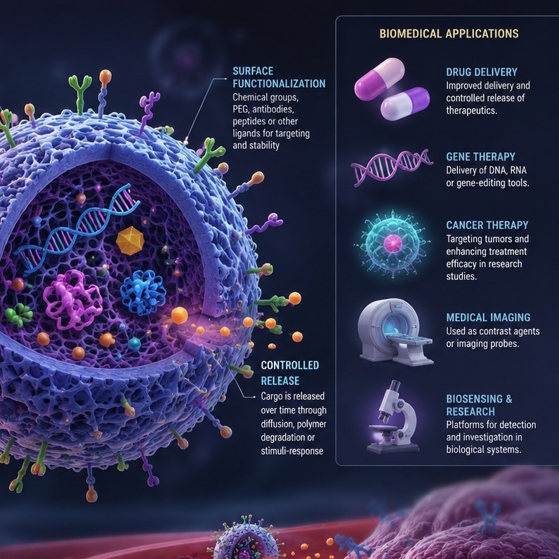
Lipid nanoparticles (LNPs) have become one of the most important nanomedicine platforms for the delivery of therapeutic molecules. Their ability to encapsulate and protect sensitive biological cargo, facilitate cellular uptake and, in appropriately designed formulations, promote intracellular release has made them particularly valuable for delivering nucleic acids such as messenger RNA (mRNA) and small interfering RNA (siRNA).

The clinical success of lipid nanoparticle-based medicines has demonstrated the potential of nanotechnology to overcome some of the limitations associated with conventional drug delivery. A major milestone was the development of LNP formulations for RNA therapeutics, including the siRNA medicine patisiran and LNP-formulated mRNA COVID-19 vaccines. These applications have also accelerated research into LNPs for gene editing, cancer treatment, protein replacement, vaccination, imaging and other biomedical applications.

At the same time, LNP technology continues to evolve. Researchers are developing new lipid chemistries, manufacturing strategies and targeting approaches to improve tissue specificity, stability, safety and therapeutic efficacy.

What are Lipid Nanoparticles?

Lipid nanoparticles are nanoscale delivery systems composed primarily of lipids that self-assemble into particles capable of carrying therapeutic or diagnostic molecules.

Although the terms are sometimes used interchangeably, lipid nanoparticles and liposomes are not the same technology. Liposomes generally contain an aqueous compartment enclosed by one or more lipid bilayers, whereas many modern LNP formulations used for nucleic acid delivery have more complex, formulation-dependent internal structures that differ from the classical bilayer organization of liposomes.

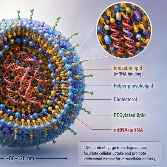
A widely used LNP architecture contains four main types of components:

  • Ionizable lipids, which interact with negatively charged nucleic acids and play a key role in particle formation, cellular uptake and endosomal escape.
  • Helper phospholipids, which contribute to particle structure and stability.
  • Cholesterol, which influences particle organization and membrane interactions.
  • PEGylated lipids, which can help control particle size, aggregation and stability.

The exact composition varies depending on the intended application. The choice of lipid chemistry and formulation parameters can strongly influence particle size, encapsulation efficiency, biodistribution, cellular uptake, endosomal escape and biological activity.

For nucleic acid delivery, LNPs are particularly useful because they can protect RNA from degradation in biological environments and facilitate its transport into cells. Following cellular uptake, successful formulations need to escape the endosomal compartment so that the cargo can reach its intracellular site of action.

Advantages of Lipid Nanoparticles

LNPs offer several properties that make them attractive for biomedical research and pharmaceutical development.

Protection of sensitive biomolecules

RNA and other biological molecules can be vulnerable to enzymatic degradation. Encapsulation within an LNP can protect the cargo during formulation, administration and transport through biological environments.

Facilitated intracellular delivery

LNPs can facilitate cellular uptake through endocytic pathways. For RNA therapeutics, the ability of appropriately designed ionizable lipids to promote endosomal escape is particularly important for achieving intracellular delivery.

Versatile cargo loading

LNPs can be engineered to carry different types of therapeutic cargo, including mRNA, siRNA and other nucleic acids. They can also be investigated for the delivery of proteins, small-molecule drugs and combinations of therapeutic agents.

Tunable physicochemical properties

Particle size, lipid composition, surface characteristics, cargo-to-lipid ratio and other formulation parameters can be modified to influence biological behaviour. This tunability provides opportunities to adapt LNPs to different therapeutic objectives.

Established clinical translation for nucleic-acid delivery

Unlike many nanomedicine platforms that remain primarily at the preclinical stage, LNP technology has already demonstrated clinical utility for nucleic-acid delivery. Patisiran uses an LNP formulation to deliver siRNA to the liver, while LNPs are also used to deliver mRNA in COVID-19 vaccines.

Potential for scalable manufacturing

Modern LNP production frequently uses controlled rapid mixing processes, including microfluidic and related mixing technologies. These approaches can provide reproducible particle formation and offer potential pathways for scalable manufacturing.

However, LNPs are not universally optimal for every application. Challenges include tissue-specific targeting, stability, manufacturing consistency, immune responses and potential toxicity associated with particular lipid compositions. Current research therefore focuses on developing increasingly selective and biocompatible formulations.

How are Lipid Nanoparticles Synthesized?

The preparation of LNPs depends on their composition and intended application. For nucleic acid-loaded LNPs, one of the most widely used approaches is rapid mixing of a lipid-containing organic phase with an aqueous phase containing the nucleic acid.

Typically, the lipids are dissolved in an organic solvent, commonly ethanol, while the nucleic acid is prepared in an aqueous buffer under appropriate pH conditions. When the two streams are rapidly mixed, changes in solvent composition and lipid ionization promote lipid self-assembly and nanoparticle formation. The nucleic acid becomes associated with or encapsulated within the developing particles.

Microfluidic mixing

Microfluidic systems can provide highly controlled mixing conditions and are particularly attractive for LNP formulation development. Parameters such as flow rates, mixing conditions, lipid concentration and aqueous-to-organic phase ratios can affect particle size, polydispersity and encapsulation efficiency.

Following particle formation, additional processing steps may be required. These can include removal or reduction of organic solvent, buffer exchange, concentration and sterile filtration, depending on the formulation and intended application.

Characterization

A comprehensive characterization strategy is essential to determine whether an LNP formulation has the required properties. Common parameters include:

  • Particle size and size distribution
  • Polydispersity
  • Surface characteristics
  • Encapsulation efficiency
  • Lipid composition and purity
  • Cargo integrity
  • Stability during storage
  • In vitro cellular uptake and biological activity
  • In vivo biodistribution and pharmacokinetics, when appropriate

For therapeutic development, formulation characterization must ultimately be connected to critical quality attributes and biological performance.

Biomedical Applications of Lipid Nanoparticles

LNPs are being investigated across a broad range of biomedical applications. Some applications, particularly nucleic acid delivery, have already reached clinical use, whereas others remain at the research or preclinical development stage.

1. Biomolecule Delivery and RNA Therapeutics

One of the most important applications of LNPs is the delivery of biomolecules and nucleic acids.

Nucleic acids such as mRNA and siRNA offer powerful therapeutic possibilities but face important delivery challenges. They are relatively large, hydrophilic and susceptible to degradation, making it difficult for them to cross cellular membranes efficiently.

LNPs can address several of these barriers by encapsulating the nucleic acid, protecting it from degradation and promoting cellular uptake. Once inside the cell, the objective is to release the cargo from the endosomal compartment so that it can exert its biological function.

mRNA delivery

LNPs have become particularly important for mRNA delivery. Once delivered to the cytoplasm, mRNA can be translated by the cell into the encoded protein without needing to enter the nucleus or integrate into the genome.

This approach has been used clinically in mRNA vaccines and is being investigated for applications including:

  • Infectious disease vaccination
  • Cancer immunotherapy
  • Protein replacement
  • Genetic and metabolic disorders
  • Regenerative medicine
  • Genome-editing approaches

The success of LNP-mRNA vaccines demonstrated that this delivery strategy can be translated from laboratory research into large-scale clinical applications.

siRNA delivery and gene silencing

LNPs can also deliver small interfering RNA (siRNA). siRNA can trigger sequence-specific degradation of complementary messenger RNA, reducing the production of a target protein.

A prominent clinical example is patisiran, an LNP-based siRNA therapy that targets transthyretin (TTR) mRNA in the liver. Its development provided important proof of concept for LNP-mediated RNA interference in patients.

Genome editing

LNPs are also being explored as delivery systems for genome-editing technologies, including CRISPR-based systems. In these approaches, LNPs can potentially deliver combinations of nucleic acids encoding or constituting components required for genome editing.

Most LNP-based in vivo genome-editing applications remain investigational, with major challenges around tissue targeting, delivery efficiency, safety and control of editing activity.

2. Imaging and Biodistribution

LNPs can also be investigated as nanocarriers for imaging agents.

By incorporating or associating imaging probes with lipid nanoparticles, researchers can study the biodistribution, cellular uptake and biological behaviour of nanoparticles in vivo. Imaging can therefore be useful not only for diagnosis but also for understanding how a formulation behaves after administration.

For example, imaging techniques can help evaluate the pharmacokinetics and biodistribution of RNA-loaded LNPs during preclinical development. This information can be valuable when optimizing formulations and identifying strategies for improving tissue targeting.

LNPs may also be designed as multifunctional platforms combining diagnostic and therapeutic functions, an approach often referred to as theranostics. However, these applications remain under investigation rather than being established clinical technologies.

3. Regenerative Medicine

LNPs are increasingly being investigated in regenerative medicine, particularly as delivery systems for nucleic acids that can modulate cellular behaviour.

mRNA-loaded LNPs, for example, can transiently provide cells with instructions to produce specific proteins. This creates opportunities to investigate protein expression involved in tissue repair, cell differentiation, angiogenesis and other regenerative processes.

Potential applications include tissue engineering, wound healing, cardiovascular regeneration and the development of strategies for directing cell fate.

Importantly, much of this work remains preclinical. The ability to control where and when therapeutic mRNA is expressed remains one of the major challenges for translating these approaches into regenerative medicine.

4. Controlled Drug Release

LNPs can be engineered to act as controlled drug delivery systems, potentially modifying the pharmacokinetics of therapeutic compounds and influencing where and when cargo becomes available.

Lipid composition, particle structure, cargo properties and environmental conditions can affect release behaviour.

This approach is being investigated for the delivery of small-molecule drugs as well as biological therapeutics. By modifying the formulation, researchers can aim to improve drug solubility, protect sensitive compounds and alter their distribution within the body.

The precise release profile, however, depends strongly on the specific LNP formulation and cargo, meaning that controlled release should be considered a design objective rather than an inherent property of every LNP.

5. Targeted Therapies

One of the major objectives in LNP research is the development of targeted drug delivery systems.

Many conventional LNP formulations exhibit substantial uptake by the liver and by cells of the mononuclear phagocyte system. Consequently, researchers are developing new lipid structures, surface modifications, ligands and formulation strategies to improve delivery to specific organs, tissues or cell populations.

Active targeting strategies may involve incorporating molecules capable of interacting with receptors expressed by particular cell types. Other approaches seek to exploit differences in physiology, biodistribution or protein adsorption to influence where nanoparticles accumulate.

Improving tissue-specific delivery is particularly important for applications such as cancer therapy, neurological disorders and gene editing, where delivery to the appropriate target cells can determine therapeutic success. However, achieving reliable tissue- and cell-specific delivery in vivo remains a major challenge, particularly outside organs such as the liver where many LNP formulations naturally accumulate.

Frequently Asked Questions

What are lipid nanoparticles used for?

Lipid nanoparticles are used to deliver therapeutic and biological molecules, particularly nucleic acids such as mRNA and siRNA. They are also being investigated for drug delivery, vaccines, gene editing, imaging, regenerative medicine and targeted therapies.

Are lipid nanoparticles the same as liposomes?

No. Although both are lipid-based nanocarriers, their structures and compositions differ. Liposomes generally contain an aqueous compartment surrounded by lipid bilayers, whereas many modern LNPs used for nucleic acid delivery have more complex, formulation-dependent internal structures and are specifically engineered for efficient nucleic acid encapsulation and intracellular delivery.

What are lipid nanoparticles made of?

Many nucleic acid LNPs contain an ionizable lipid, a helper phospholipid, cholesterol and a PEGylated lipid. However, the composition varies between formulations and applications.

How do lipid nanoparticles deliver mRNA?

LNPs protect mRNA and facilitate its uptake by cells. Following cellular internalization, the formulation must promote endosomal escape so that the mRNA can reach the cytoplasm, where it can be translated into protein.

Have lipid nanoparticle-based medicines been clinically approved?

Yes. LNP technology has reached clinical use. Patisiran is an LNP-based siRNA medicine, while LNPs are also used to deliver mRNA in COVID-19 vaccines. These applications have provided important clinical validation of LNP-based nucleic acid delivery.

Can lipid nanoparticles be targeted to specific tissues?

Potentially, yes. Researchers are developing lipid chemistries, surface modifications and other formulation strategies to improve tissue and cell targeting. However, achieving reliable delivery to specific tissues remains a major challenge, particularly for extrahepatic tissues where many LNP formulations do not naturally accumulate efficiently.

Are lipid nanoparticles safe?

Safety depends on the specific formulation, cargo, dose, administration route and intended application. LNPs have demonstrated clinical utility, but their lipid components and biodistribution can influence immune responses and other biological effects. Consequently, safety and tolerability must be evaluated for each formulation rather than assumed from the performance of another LNP.

Conclusion

Lipid nanoparticles have evolved from an experimental nanomedicine technology into a clinically validated platform for certain types of nucleic-acid delivery. Their clinical use in siRNA medicines and mRNA vaccines demonstrates the potential of engineered lipid systems to overcome important biological barriers to therapeutic delivery.

At the same time, the field is moving beyond these established applications. Researchers are developing LNPs for gene editing, protein replacement, cancer immunotherapy, targeted drug delivery, diagnostic imaging and regenerative medicine. New lipid chemistries and manufacturing technologies are also being developed to improve tissue specificity, intracellular delivery, stability and safety.

For biomedical research and pharmaceutical development, the ability to tailor LNP composition and physicochemical properties provides a versatile platform for designing advanced delivery systems. Continued progress in formulation, characterization, biological validation and manufacturing will be essential for expanding LNP applications beyond their current clinical uses.

Related articles

  • ...

What is NANBIOSIS?

The goal of NANBIOSIS is to provide comprehensive and integrated advanced solutions for companies and research institutions in biomedical applications. All of this is done through a single-entry point, involving the design and production of biomaterials, nanomaterials, and their nanoconjugates. This includes their characterization from physical-chemical, functional, toxicological, and biological perspectives (preclinical validation).

Leading scientists

The main value of NANBIOSIS is our highly qualified and experienced academic scientists, working in public institutions, renowned universities and other research institutes.

Custom solutions

Designed for either scientific collaboration or the private industry, we adapt our services to your needs, filling the gaps and paving the way towards the next breakthrough.

Cutting-Edge facilities

Publicly funded, with the most advanced equipment, offering a wide variety of services from synthesis of nanoparticles and medical devices, including up to preclinical trials.

Standards of quality

Our services have standards of quality required in the pharmaceutical, biotech and medtech sectors, from Good Practices to ISO certifications.

In order to access our Cutting-Edge Biomedical Solutions with priority access, enter our Competitive Call here.

NANBIOSIS has worked with pharmaceutical companies of all sizes in the areas of drug delivery, biomaterials and regenerative medicine. Here are a few of them:
Read More

NANBIOSIS Supports Scientific Outreach at the Launch of Misión Aragón: Cracks de la Ciencia

NANBIOSIS joined the launch of Misión Aragón: Cracks de la Ciencia, promoting scientific outreach and inspiring future generations through Aragón’s research excellence.

Zaragoza, July 2026 — NANBIOSIS took part in the presentation of Misión Aragón: Cracks de la Ciencia, a new science outreach initiative promoted by the Government of Aragón to inspire scientific vocations among young people by celebrating the region’s most influential researchers and world-class scientific infrastructures.

The initiative, presented on 30 July 2026 at the Pablo Serrano Museum in Zaragoza, introduces a collection of 15 collectible cards featuring ten outstanding Aragonese scientists and the five Unique Scientific and Technical Infrastructures (ICTS) located in Aragón, including NANBIOSIS. Through an engaging, game-based format, the project encourages children and families to discover the people, institutions and facilities that are shaping scientific progress while exploring the region of Aragón.

Representatives of the institutions and family members from the scientists.

Representing CIBER-BBN and NANBIOSIS, Gabriel Alfranca, Internationalization and Communication Manager, attended the event and was interviewed about the importance of scientific infrastructures and communication in bringing research closer to society. His participation highlighted the role that research platforms such as NANBIOSIS play in supporting biomedical innovation and making cutting-edge science accessible beyond the laboratory.

Among the featured cards is CIBER-BBN, which can be collected at La Alfranca (Zaragoza) alongside the card dedicated to scientist Martina Bescós. By including leading research infrastructures in the collection, the initiative showcases the scientific ecosystem that enables discoveries with societal impact and encourages younger generations to engage with science and technology.

The event also acknowledged the recent National Research Award Juan de la Cierva for Knowledge Transfer received by Jesús Martínez de la Fuente, Research Professor at CSIC, researcher at INMA (CSIC–University of Zaragoza), and one of the Coordinators of CIBER-BBN. The award recognizes his outstanding career in nanobiotechnology and his exceptional contribution to transforming scientific research into real-world biomedical applications, reinforcing Aragón’s position as a hub for scientific excellence and innovation.

Prof. Jesús M. de la Fuente

Through initiatives such as Misión Aragón: Cracks de la Ciencia, public institutions, research centres and scientific infrastructures are working together to promote scientific culture, foster STEAM vocations and inspire the next generation of researchers by demonstrating that science is both accessible and capable of transforming society.

What is NANBIOSIS?

The goal of NANBIOSIS is to provide comprehensive and integrated advanced solutions for companies and research institutions in biomedical applications. All of this is done through a single-entry point, involving the design and production of biomaterials, nanomaterials, and their nanoconjugates. This includes their characterization from physical-chemical, functional, toxicological, and biological perspectives (preclinical validation).

If you want to collaborate with us, visit our Order Request page.

Leading scientists

The main value of NANBIOSIS is our highly qualified and experienced academic scientists, working in public institutions, renowned universities and other research institutes.

Custom solutions

Designed for either scientific collaboration or the private industry, we adapt our services to your needs, filling the gaps and paving the way towards the next breakthrough.

Cutting-Edge facilities

Publicly funded, with the most advanced equipment, offering a wide variety of services from synthesis of nanoparticles and medical devices, including up to preclinical trials.

Standards of quality

Our services have standards of quality required in the pharmaceutical, biotech and medtech sectors, from Good Practices to ISO certifications.

In order to access our Cutting-Edge Biomedical Solutions with priority access, enter our Competitive Call here.

NANBIOSIS has worked with pharmaceutical companies of all sizes in the areas of drug delivery, biomaterials and regenerative medicine. Here are a few of them:
Read More