+34 620 10 75 37info@nanbiosis.com

Nanbiosis

New look of Nb4D – CAbS (NANBIOSIS U2) “Revolutionising Diagnosis”

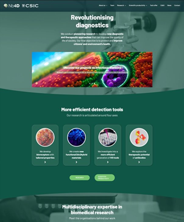
Nb4D has a new look on line! Nb4D Group (of IQAC-CSIC and CIBER-BBN) has lunched a new website “Revolutionsising Diagnosis with the aim to facilitate a faster and easier navigation througth their “pioneering research to develop new diagnostic and therapeutic approaches” and their solutions and expertise to help researchers and companies.

Antibodies, bioreceptors, hapten design and synthesis, immunoanalytical method development, new ivd tools, surface functionalization, therapeutic antibodies and much more knowledge and expertise revolutionising diagnosis.

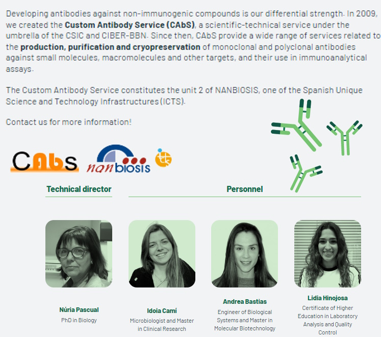
The new website contains a page for CAbS-NANBIOSIS. Custom Antibody Service (CAbS), unit 2 of the ICTS NANBIOSIS

Read More

NABIHEAL project launches website

The website for NABIHEAL, an EU-funded Horizon Europe project developing biomaterials for complex wound healing, is now online.

The Horizon Europe project NABIHEALi project is coordinated by the Center for Biomedical Research Network (CIBER) at the Institute of Materials Science of Barcelona (ICMAB-CSIC).

This project will apply one the Cutting Edge Biomedical Solutions” of NANBIOSIS for the preparation of different nanoestructures with antimicrobial properties, required for the development of the final multifunctional wound healing biomaterials. This case will gather the expertise of two NANBIOSIS unit: NANBIOSIS U6 will produce and characterize these nanoestructures with antimicrobial properties, which will be tested in NANBIOSIS U16.

Find out more about the project and what its impact will be, and browse the 14 partners from 7 countries to see how each contributes to the project’s objectives. NABIHEAL WEBSITE

Related news: New European Project NABIHEAL in biomaterials for complex wound healing

Read More

U6-E08. Mastersizer 2000

Mastersizer 2000 – Malvern

Description:

The Mastersizer 2000 uses the laser diffraction technique to measure the size and particle size distribution of materials. This is carried out by measuring the intensity of light scattered when a laser beam passes through a sample of dispersed particles. The detectors measure light intensity for red and blue light. The data is analyzed to calculate the size of the particles that created the scattering pattern.

The equipment contains two sample dispersion units:
– Wet: for samples in solution, which can be applicable to different solvents.
– Dry: for solid samples in powder form, which are dispersed by a current of air.

Technical specifications:

• Particle size: 10 nm – 3.5 mm
• Measurement principle: Mie and Fraunhofer dispersion
• Light source:
– Red: He-Ne (632.8 nm)
– Blue: LED (470 nm)
• Optical Alignment: Automatic rapid alignment system with darkfield optical reticle and multi-element alignment detector
• Dispersion units:
– Dry route
– Wet route
• Operating temperature: 5º to 40º C

Applications:

• Molecular and particle size distribution analysis.
• Powder fluidity analysis
• Particle compression density analysis
• Stability analysis in suspensions and emulsions
• Analysis of the dissolution rate of materials
• Determination of the reaction rate in solid systems

Read More

U6-E10. Nanosight NS-300 for Nanoparticle Tracking Analysis by fluorescence mode.

NanoSight NS300 – Malvern

Description:

The NanoSight NS300 uses nanoparticle tracking analysis (NTA) technology, which uses the properties of light scattering and Brownian motion to obtain measurements of concentration and size distribution of particles in liquid suspension.
A laser beam passes through the sample chamber, and suspended particles in the path of this beam scatter the light in such a way that they can be easily seen through a 20x microscope on which a camera is mounted. The camera runs at 30 frames per second and captures a video of the particles in their natural Brownian motion. The software tracks the particles individually and, using the Stokes-Einstein equation, calculates the hydrodynamic diameter of each of them.

Technical specifications:

• Size range: 10 – 1000 nm
• Concentration range: 106 – 109 particles/ml
• Temperature control range: 5º C below ambient to 50º C
• Minimum sample volume: 250 µl
• Camera: SCMOS
• Focus: Computer controlled motorized focus
• Fluorescence: 6-place motorized filter wheel, with filter options
• Wavelength:
– Blue: 488nm
– Green: 532nm

Applications:

• Molecular and particle size distribution analysis.
• Concentration measurement.

Update of the Nanosight system

  1. The Green laser module for the NanoSight NS300 equipment.

The NS300 allows analysis of the size distributionand concentration of all types of nanoparticles from 0.01 – 1 µm in diameter. This new laser module will allow NTA to measure a range of fluorescent particles, avoiding interferences during the measurements due to sample (auto)fluorescence and absorption.  This is done by detecting the fluorescence signal, which is emitted naturally by particles or as a result of fluorescence labeling or tagging.

This image has an empty alt attribute; its file name is image-10-1024x768.png

Financed by the project FICTS-1420-27

         2.  Nanosight NS300 Violet Laser Module 405nm

Description: The NanoSight NS300 instrument provides a reproducible and easy-to-use platform for nanoparticle characterization. The NS300 allows for quick and automatic analysis of size distribution and concentration of all sorts of nanoparticles, ranging from 10 to 1000 nm in diameter, depending on instrument configuration and sample type. Thanks to the possibility of adding interchangeable laser modules and the introduction of a 6-position motorized filter wheel, analysis of different fluorophores can be performed. The sample temperature is fully programmable through the nanoparticle tracking analysis (NTA) software.

Technical Specifications:

  • Size Range: 10 nm-1000 nm
  • Concentration Range: 10^6-10^9 particles/mL
  • Minimum Sample Volume: 250 µL
  • Laser Type: 405 nm violet laser
  • Fluorescence Filter: 430 nm
  • Temperature Range: 5 ºC below ambient temperature up to 70 ºC

Applications:  The equipment can be used in various applications and to characterize different types of particles, such as: Extracellular vesicles, Viruses and vaccines, Drug delivery, Biotherapeutics, Colloids and Nanomaterials, Ultrafine bubbles.

This image has an empty alt attribute; its file name is Láser-violeta-1024x768.jpg

Financed by the project ICT2021-006987

Read More

U6-E17. Surface Plasmon Resonance (SPR) system

SR7000 DC Reichert

Description: Dual Channel Modular platform, outstanding value. This Surface Plasmon Resonance (SPR) system generates high-quality data for kinetics, affinity, thermodynamics, and concentration. The SR7000DC SPR System provides SPR technology for label-free biomolecular interaction analysis.

Reichert’s SR7000DC SPR System is an innovative label-free detection system offering unparalleled flexibility and remarkable sensitivity for biomolecular and biochemical analysis. Designed for breakthrough discoveries, it is component-based and cost-effective; upgrade or add instruments at any time. The system is low maintenance, using off-the-shelf fluidics.

 Technical specifications:

Measurement Channels: Two

Sample Loading: Autosampler. Up to 768 samples

Injection Volume: 1 uL to 4,500 uL (depends on installed loop volume)

Buffer Degasser: Built-in

Temperature Range: 10 °C below ambient to 70 °C

Sample Storage: 4 °C or ambient temperature

21 CFR Part 11: Compliant

Measurement sensitivity

Baseline Noise: 0.05 µRIU (RMS)

Baseline Drift: < 0.1 µRIU/min

Minimum Molecular Weight Detection: No lower limit for organic molecules

Typical kinetic and equilibrium constant ranges

Association Rate Constant: 103 to 108 M-1 sec-1

Dissociation Rate Constant: 10-1 to 10-6 sec-1

Equilibrium Dissociation Constant: 10-3 to 10-12 M

 Applications: SPR is typically used in academic and industry settings to advance research in a range of areas, including:

  • Antibody development.
  • Drug development and discovery.
  • Concentration analysis.
  • Gene regulation.
  • Nanoparticles/Nanomaterials.
  • Pharmaceutical research.
  • Studying protein structure and function determination.
  • Systems biology.
  • Thermodynamics analysis (ΔH & ΔS).

Whether you’re studying Alzheimer’s disease, developing new vaccines or focused on other areas of protein research, SPR can help you quickly determine whether proteins interact, and which ones are worth studying more.

Read More

U6-E18. DOE

Design of experiments (DOE) software

Description: Software platform and assistance for performing Design of Experiments (DOE), a systematic and statistical approach to planning, conducting, and analyzing experiments or tests. It involves strategically manipulating variables to gather relevant and reliable information while minimizing resources and time.

Applications: DOE helps researchers and engineers understand the relationships between factors, identify optimal conditions, and improve processes by optimizing performance and reducing variability. Applicable in most fields of production and research.

Read More

Safe-n-MedTech celebrates its 48th Month Management Committee Meeting

The event brought together high-level experts from 3 continents in the field of nanotechnology.

Barcelona, March 27th, 2023

The EU funded project Safe-n-MedTech celebrated its official 48th Month Management Committee Meeting on March 7th to 9th 2023 with a private event taking place in Lisbon. The event was organized and hosted by Instituto Medicina Molecular João Lobo Antunes (IMM), who are partners in this project.

The Consortium had the pleasure to count with the virtual attendance of the Project Adviser, Ozlem Cangar (HADEA), who could follow the critical aspects of the project progress.

The meeting was opened by Ángel del Pozo, Deputy Manager of Programs Strategy at Biokeralty Research Institute, and Scientific and Technical Manager of the project, who offered a comprehensive overview of the latest developments concerning each of the work packages.

Following this, work package leaders and representatives from all partners presented the most significant advances in their current tasks, and highlighted the results obtained to date in each of the project’s work packages. They also had the opportunity to share the latest progress within the Test Cases that are being carried out. On the second day, the consortium established the critical tasks and timelines to fulfill for the successful completion of the project.

Prior to the main event, a business meeting was held at the facilities of IMM with the aim to discuss the next steps for the successful development of the OITB Pathway, a non-profit organization derived from this project, which will act as a single-entry point (SEP) to help technologies in health care into the market.

With less than 7 months ahead towards the end of the project it is expected that work will continue in a collaborative and committed manner to achieve the established objectives and goals. At this stage, partners are strongly focused on public relations and communication activities. Dissemination of project results is essential for long-term success.

The two and a half-day event offered all partners the opportunity to discuss about all the ongoing activities, next challenges and the best way to face them. “The project ends in 7 months, but we will continue building OITB Pathway and delivering all our services and knowledge. We have set the path with clear plans towards the future!”, Ángel del Pozo pointed.

About Safe-n-MedTech
The EU funded Safe-n-MedTech is a 4-year project that is working on an open access, innovative platform to provide the knowledge, networks and services needed for the development of nanotechnology-based medical and diagnostic devices. The objective is to offer the service to leading companies and laboratories.

For more information visit: www.safenmt.com

Read More

NANBIOSIS U16 enlarges its capacities with a new FlexPS-ARPES-E kit

NANBIOSIS unit 16 Surface Characterization and Calorimetry Unit form CIBER-BBN and University of Extremadura has recently incorporated new equipment acquired through the execution of the project FICTS1420-14-09, cofinanced with FEDER funds, the Ministry of Economy and Competitiveness and Junta de Extremadura, Regional Ministry of Economy, Science and Digital Agency. A new FlexPS-ARPES-E kit, manufactured by SPECS, is now available for use by researchers

This new resource uses the technique of photoelectron spectroscopy generated by X-rays to analyze the chemical composition of a surface. The electrons given off by the irradiated surface generate a fingerprint of the molecules that make up that surface, yielding information about the chemical composition of approximately the first 10 nm (one millionth of a millimeter) of thickness, making it a technique of Last generation.

One of the advantages of the acquired equipment is that the type of analyzer it has allows for different configurations to obtain different measurement modes. Thus, depending on the application, you can choose between:

XPS/ESCA (X-ray Photoelectron Spectroscopy) mode: The excitation source is X-ray.

SEM/SAM (Scanning Electron Microscopy/Auger) mode: the excitation source is electrons.

UPS mode (Ultraviolet Ray Photoelectron Spectroscopy): The excitation source is UV rays.

ISS (Ion Scattering Spectroscopy) mode: the excitation source is ions. This excitation source can also be used for depth profiling.

At a cost of 800,000 euros, its acquisition has been achieved thanks to financial support from the Ministry of Science and Innovation, the General Secretariat for Research and FEDER funds from the multi-regional operational program of Spain in the line of action of Singular Scientific and Technical Infrastructures ( ICTS). In addition, it has been co-financed by the Junta de Extremadura, the Ministry of Economy, Science and Digital Agenda and the General Secretariat of Science, Technology, Innovation and University.

Other equipment financed in this same action is:

A DMC8 Leica profilometer: a device that allows determining the 3D texture of surfaces through spatial, volumetric and height parameters, from the millimeter to the nanometer range.

A Krüss DSA100E/ DSA100M goniometry : system for determining the surface tension of solids and liquids. The equipment has a microdrop dosing system, a thermostatic chamber and a chamber for controlling the vapor saturation of the liquids being analysed. In addition, the microdroplet system has a tilting base that allows the samples to be tilted by at least 90°.

An additional cannon for a team of TOF-SIMs: a team of secondary ion mass spectrometry by time of flight (TOF-SIMS), a very sensitive technique for analyzing the composition of surfaces that provides detailed elemental and molecular information of coatings, layers fine lines and interfaces both at the superficial and three-dimensional level.

Read More

A discovery in one of the most aggressive cancers will allow more efficient diagnosis

The extracellular vesicles secreted by triple-negative breast cancer stem cells are markers of lung metastasis, according to a study carried out by researchers at CIBER.

The work has been carried out by researchers from various CIBER-BBN groups (Bioengineering, Biomaterials and Nanomedicia), and CIBERONC (CIBER area focused on cancer) has participated in it. The research has been led by Joaquín Seras, from the Vall d’Hebron Research Institute (VHIR), a specialist in targeted drug therapies.

Physicochemical EVs characterization and all the in vivo studies were performed by NANBIOSISunits of CIBER, specifically NTA analysis was carried out at Unit 6 of Biomaterial Processing and Nanostructuring, led by Nora Ventosa at ICMB-CSIC and animal experimentation at Unit 20 “In vivo experimental platform”, led by Ibane Abasolo at VHIR.

The vesicle, in cell biology, is an organelle that forms a small, closed compartment, separated from the cytoplasm by a lipid bilayer just like the cell membrane. The vesicles store, transport or digest cellular products and waste. According to Joaquin Seras, leader of the research: “the identification of this subpopulation of cancerous extracellular vesicles, and their important role in the progression of the disease, will allow in the future to develop systems more effective and less invasive diagnostic methods based on their detection directly from blood samples”.

In different types of tumors, including triple negative breast cancer, it has been observed that the extracellular vesicles generated by tumor cells play an important role in the generation of pre-metastatic niches. Triple negative breast cancer, one of the most aggressive, highly plastic and heterogeneous, is characterized by a significant presence of malignant stem cells.

The study carried out by the Spanish researchers from CIBER with promising results, published in the “International Journal of Cancer”, shows, both in in vitro and in vivo models of the disease, that the vesicles actively contribute to the formation of areas with favorable conditions for the formation of metastases, thus favoring way, the spread of the disease.

Research contributions
In the opinion of Joaquin Seras, the great contribution of this work is that it “describes how the extracellular vesicles secreted by certain subpopulations of cancer cells, specifically those derived from cancer stem cells, have the potential to modify the microenvironment of the future metastatic niche to promote tumor growth.

In other words, continues the leader of the study: “the research sheds new information on the pathogenic mechanism of the disease, and suggests these extracellular vesicles as markers with diagnostic potential. It should be noted that these nanoparticles are secreted into the bloodstream by tumor cells, and effective capture and identification would allow them to be exploited as a diagnostic tool”.

On the characterization of extracellular vesicles of cancer cells
The complex composition and functional differentiation of cancer cells in a tumor also increases the heterogeneity of the subsets of vesicles secreted by cancer.

This phenomenon is particularly relevant in triple negative breast cancer, one of the most aggressive, highly plastic and heterogeneous cancers, characterized by a significant presence of malignant stem cells. However, until now the diversity of the vesicles secreted by cancer cells had not been studied, a diversity that is closely related, in turn and as the study shows, to cellular heterogeneity in triple-negative tumors.

The importance of the CIBER study lies at this point: the vesicles secreted by different tumor subpopulations and grouped by their degree of differentiation show fundamentally different activities in terms of their impact on cancer progression.

In the investigation, the extracellular vesicles secreted by up to three different types of neoplastic cells have been isolated and characterized, observing different bioburdens for each type, with the consequent differential effect on stromal cells. In addition, and as the study shows, cancer stem cell-derived vesicles contribute to converting healthy lung cells into receptive niches for the metastatic growth of cancerous breast cells.

Article reference:

González-Callejo P, Gener P, Díaz-Riascos ZV, Conti S, Cámara-Sánchez P, Riera R, Mancilla S, García-Gabilondo M, Peg V, Arango D, Rosell A, Labernadie A, Trepat X, Albertazzi L, Schwartz S Jr, Seras-Franzoso J, Abasolo I. Extracellular vesicles secreted by triple-negative breast cancer stem cells trigger premetastatic niche remodeling and metastatic growth in the lungs. Int J Cancer. 2023 Jan 27. doi: 10.1002/ijc.34447. Epub ahead of print. PMID: 36705298.

Read More

Nanoparticles to modulate topography and ligand distribution at the nanoscale: impact on cell behavior

Doctor Marc Martínez from the Nanomol-Bio group – NANBIOSIS U6 from CIBER-BBN at ICMAB-CSIC, defended his PhD thesis “Nanoparticles to modulate topography and ligand distribution at the nanoscale: impact on cell behavior” on 9 March 2023 at ICMAB.

he PhD thesis was supervised by Imma Ratera, Judith Guasch and Nora Ventosa from the Nanomol-Bio group at ICMAB-CSIC.

Ana Paula Candiota Silveira, Scientific Coordinator of NANBIOSIS U25 was part of the Committee that evaluated the Thesis tooghether with Jesús Martínez de la Fuente, Instituto de Nanociencia y Materiales de Aragón (INMA-CSIC) (President),  and Anna Lagunas Targarona, Institut de Bioenginyeria de Catalunya (IBEC) (Vocal).

As Marc Martínez explained in an inteview to ICMAB “I produce nanoparticles in the lab and I use them to see how cells react to them. I work at the interface between cells and materials. My research can be applied to cell culture, which can be relevant for the development of new therapies for the regeneration of organs or for building implants to replace missing organs and body parts”.

Doctor Marc Martínez’s PhD thesis was part of the PhD Programme in Biochemistry, Molecular Biology and Biomedicine from the Universitat Autònoma de Barcelona (UAB).

Further information at ICMAB webpage

Read More