+34 620 10 75 37info@nanbiosis.com


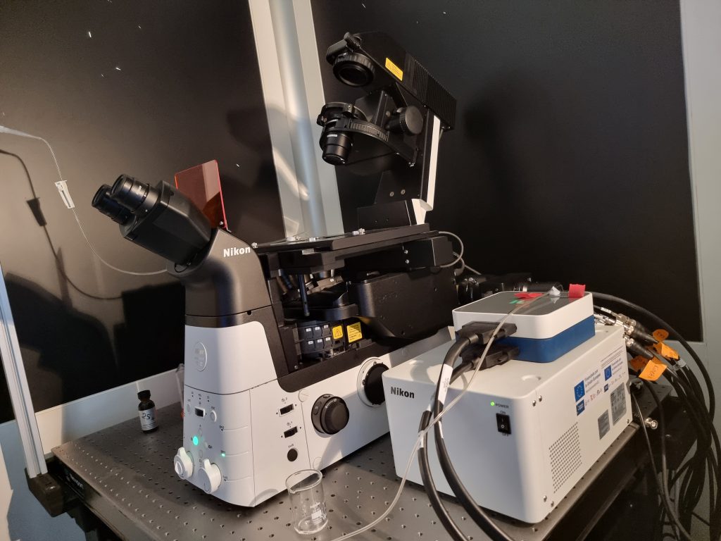
The new Nikon Eclipse Ti2-E imaging system is based on an inverted, fully motorized, large field-of-view wide-field fluorescence microscope equipped with state-of-the-art features for high-speed visualization of large tissues or sample sets. The microscope is equipped for imaging standard slides, culture dishes, multiwells, and microplates (e.g., 96-well plates). The system includes “Perfect Focus” for applications requiring outstanding focus control (e.g., image tiling), a high-resolution motorized stage, and NIS-Elements Advanced Research (AR) software with AI-based modules for customizing sample visualization and data analysis. The heart of the system is the outstanding Kinetix camera from Photometics. The back-illuminated sCMOS sensor of the camera delivers the fastest speed (up to 500 fps at 3200×3200 pixels), and the largest field of view (25 mm of FOV) with outstanding light sensitivity, significantly improving data throughput.
The camera is coupled to a high-speed illumination system to quickly switch (<50 ms) between different wavelengths, allowing fast multi-color imaging of large samples or the quantitative determination of biological quantities using fluorescent sensitive dyes. The Ti2’s exceptionally stable, drift-free platform is designed to meet the demands of super-stable imaging. At the same time, its unique hardware-triggering capabilities enhance even the most challenging, high-speed imaging applications. Furthermore, the Ti2’s unique, intelligent functions guide users through imaging workflows by gathering data from internal sensors, eliminating the possibility of user errors. In addition, the status of each sensor is automatically recorded during acquisition, providing quality control for imaging experiments and enhancing data reproducibility in a fraction of the time of conventional equipment.
These characteristics, in combination with Nikon’s advanced Artificial Intelligence (AI) analysis capabilities integrated into the microscope’s software, make real-time analysis and optimization of the measurements possible to ensure the best results through user-specific automatic experimental routines.
Specifications and capabilities: