+34 620 10 75 37info@nanbiosis.com

Posts Taged bioimaging

Submissions open for Special Issue of MDPI on Fluorescent Organic Nanoparticles for Bioimaging and Theragnostics

Nora Ventosa and Mariana Köber, from NANBIOSIS Unit 6 of CIBER-BBN and ICMAB-CSIC, and Judit Morlà-Folch, from the BioMedical Engineering and Imaging Institute at the Icahn School of Medicine at Mount Sinai, New York, are editors of the Special Issue of MDPI Pharmaceutics.

The Special Issue on Fluorescent Organic Nanoparticles for Bioimaging and Theragnostics belongs to the “Nanomedicine and Nanotechnology” section and has a deadline for manuscript submissions on 25 July 2022.

The guest editors explain the main topic of this Special Edition:

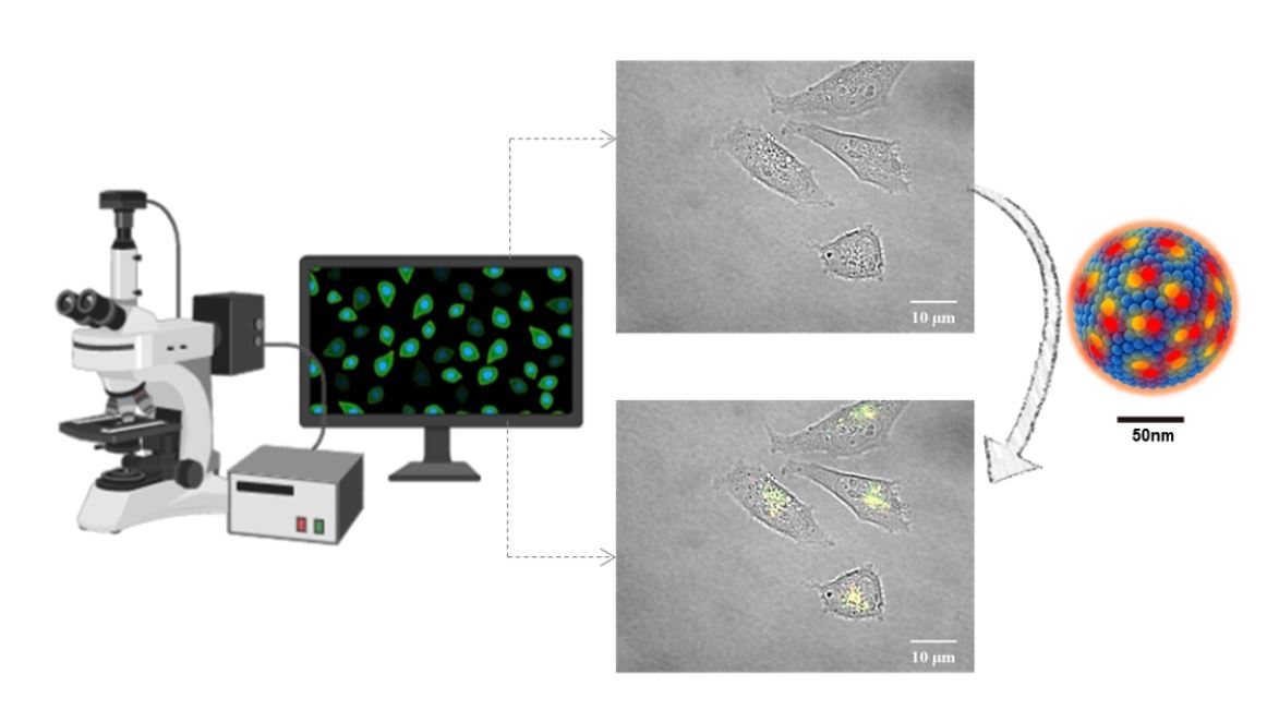
“Fluorescence-based techniques play an essential role in the study of biological events in tissues and animals due to their specificity and noninvasive nature. However, realizing the whole potential of today’s fluorescence imaging and detection in terms of speed, resolution, and sensitivity, requires fluorescent labels that combine stability, a very high brightness, and a high photostability.

In this regard, novel, bright and stable organic fluorescent nanoparticles have evolved rapidly during the last few years, allowing further development of novel, experimental treatments and imaging strategies, including photodynamic therapy or image-guided surgery.

These results shine a spotlight on fluorescent nanomaterials as promising candidates for imaging and theragnostics in several health disorders. In this Special Issue, we invite authors to report on their recently developed, fluorescent, organic nanoparticles for imaging, diagnostics, and the treatment of diseases.”

If you have a relevant manuscript, you can submit it at MDPI in the submission form before 25 July 2022. All papers will be peer-reviewed, and research articles, review articles or short communications are invited. For planned papers, a title and short abstract (about 100 words) can be sent to the Editorial Office for announcement on this website. More information on submission here

About the Pharmaceutics MDPI Journal

Pharmaceutics (ISSN 1999-4923) is an online open access journal on the science and technology of pharmaceutics and biopharmaceutics. The scientific community, the wider community and the general public have unlimited and free access to the content as soon as a paper is published; this open access to your research ensures your findings are shared with the widest possible audience. Please consider publishing your impressive work in this high quality journal. We would be pleased to welcome you as one of our authors.” – Editor-in-Chief Prof. Dr. Yvonne Perrie from the Strathclyde Institute of Pharmacy and Biomedical Sciences at the University of Strathclyde.

Source of information: ICMAB-CSIC

Read More

New fluorescent organic nanoparticles to see the invisible

A new nanomaterial for bioimaging has been developed by researchers at NANBIOSIS Unit 6 Biomaterial Processing and Nanostructuring Unit from the Nanomol group from ICMAB-CSIC and CIBER-BBN . The researchars are also members of the TECNIO technology transfer network ACCIÓ-Generalitat de Catalunya, together with the New Jersey Institute of Technology (NJIT, USA) and the University of Parma (UNIPR, Italy). The results of the study are the result of the TECNIOspring PLUS project co-financed by ACCIÓ and the European Commission.

It is true that it is very difficult to understand what happens in our bodies if we are unable to visualise it. For example, we currently know that tumour cells have the capacity to grow without control thanks to various microscopic techniques that have allowed us to enlarge them to such an extent that we have been able to see each cell perfectly. The design of microscopes and the optical and electronic engineering behind them has advanced very rapidly in recent years. In fact, the 2014 Nobel Prize in Chemistry was awarded to researchers Eric Betzig, William E. Moerner and Stefan Hell, for the development of super-resolution fluorescence microscopy. These advances have made it possible to see even what is inside cells, reaching the nanometer scale with high resolution.

Now, what happens when we are not able to see what we are looking for? This is where fluorescent probes come into play, molecules that provide a signal: they emit light at a certain wavelength once they are excited. These probes must meet a series of requirements, among which are: they must have a high luminosity or brightness, be totally biocompatible, and have high photo-stability and high dispersibility in physiological media.

The Nanomol group has developed new fluorescent probes, specifically fluorescent organic nanoparticles (FONs). These new FONs are based on Quatsomes (QSs), nanovesicules produced by the same group through a green technology (Delos-susp, Nanomol Technologies SL), which are charged with fluorophores or fluorescent molecules – specifically two types of carbocyanins. The nanoparticles have an average diameter of 120 nm and have demonstrated good biocompatibility and high stability, both over time and once exposed to high power laser irradiation.

Characterization of nanovesicles was made at the ICTS “NANBIOSIS”, more specifically by the Unit 6 Biomaterial Processing and Nanostructuring Unit of CIBER-BBN.

“The brightness achieved is especially relevant: these new fluorescent nanoparticles are about 100 times brighter than other commercial fluorescent nanoparticles, such as Quantum Dots, thus allowing the acquisition of high quality images” explains Judit Morla-Folch, postdoctoral researcher of the Nanomol group at the ICMAB and first author of the study, published in the journal ACS Appl. Mater. Interfaces.

In addition, these nanoparticles have another singularity, and that is that they experience Förster resonance energy transfer, usually abbreviated as FRET. This phenomenon allows for improved image acquisition as it significantly reduces self-absorption and therefore background noise during bioimage acquisition. In addition, the FRET effect allows the integrity of the nanoparticle to be monitored, a great advantage for biomedical applications where it is necessary to know when the nanovesicle remains as a whole or it disintegrates.

In summary, the fluorescent organic nanoparticles (FONs) developed by the Nanomol group of the ICMAB-CSIC in collaboration with the NJIT (USA) and the UNIPR (Italy) constitute a promising platform for bioimaging and for the design of medical diagnostic kits.

Cover Figure: The new fluorescent organic nanoparticles allow to improve the visualization of cells and tissues under the microscope.

Reference article:

Dye-Loaded Quatsomes Exhibiting FRET as Nanoprobes for Bioimaging
Judit Morla-Folch, Guillem Vargas-Nadal, Tinghan Zhao, Cristina Sissa, Antonio Ardizzone, Siarhei Kurhuzenkau, Mariana Köber, Mehrun Uddin, Anna Painelli, Jaume Veciana, Kevin D. Belfield, and Nora Ventosa
ACS Appl. Mater. Interfaces 2020, 12, 18, 20253–20262
DOI: 10.1021/acsami.0c03040

Read More