+34 620 10 75 37info@nanbiosis.com

Posts Taged exosomes

Gold Nanoparticles Synthesized by NANBIOSIS U9 will destroy tumor cells without drugs

A CIBER-BBN team at the University of Zaragoza has developed intelligent shuttles (cell vesicles -exosomes-) to transfer nanoparticles to the interior of tumor cells and destroy them by means of heat and without drugs, following the “Trojan horse” strategy.

NANBIOSIS U9, “Synthesis of Nanoparticles Unit has developed the procedure to be able to internalize gold nanoparticles, with surface plasmon in the NIR electromagnetic range, inside extracellular vesicles derived from stem cells. The synthesis of the gold nanoparticles has been produced according to the synthesis procedures of UNIT 9 of the ICTS NANBIOSIS based on the galvanic substitution reaction in the liquid phase of Co atoms by Au+3 ions, generating a hollow structure whose geometry gives gold nanoparticles unique optical properties that allow the absorption of NIR light and its conversion into heat” , explain the researchers of NANBOSIS U9 Pilar Martín-Duque, Victor Sebastián and Jesús Santamaría.

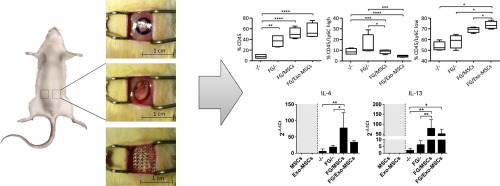
They are gold nanoparticles belonging to what is known as “plasmonic nanoparticles” that have the ability to heat up when receiving near-infrared radiation, which penetrates the body. It is, therefore, a treatment without drugs, which uses the heat generated by the particles to cause cell death around them. These particles are taken to the tumor by exosomes, having been proved efectived in animal models.

“We have managed to reduce or eliminate tumors in mice without drugs, only with the heat generated by irradiating them with a laser. In other words, we inject the exosomes with the nanoparticles into the tail of the mouse and they alone “search” for the tumor, not only in conventional models but also in multinodular ones, similar to metastatic processes”, explains Pilar Martín Duque.

For futher information:

https://www.ciber-bbn.es/noticias/desarrollan-lanzaderas-inteligentes-para-destruir-celulas-tumorales-desde-su-interior-mediante-calor-y-sin-farmacos

Article of reference:

Transfer of photothermal nanoparticles using stem cell derived small extracellular vesicles for in vivo treatment of primary and multinodular tumors. María Sancho-Albero, Miguel Encinas-Giménez, Víctor Sebastián, Estela Pérez, Lluis Luján, Jesús Santamaría, Pilar Martín-Duque Journal of Extracellular Vesicles 2022 https://onlinelibrary.wiley.com/doi/full/10.1002/jev2.12193

Read More

Mechanism and Consequences of the Impaired Hif-1α Response to Hypoxia in Human Proximal Tubular HK-2 Cells Exposed to High Glucose

NANBIOSIS has been informed about a recent publication in the pretigious scientific magzine SCIENTIFIC REPORTS (Q1) of Nature Research group, mentioning NANBIOSIS Unit 17 in the Methods section:

Immunofluorescence analysis: Detection was performed by using a Leica SP5 confocal microscope (Leica Microsystems, Wetzlar, Germany), through the Confocal Microscopy Service of the ICTS ‘NANBIOSIS’ Unit 17 of the Biomedical Research Networking Centre on Bioengineering, Biomaterials and Nanomedicine (CIBER-BBN) at the University of Alcalá, Madrid, Spain. HIF-1α-dependent immunofluorescence intensity was quantified after digital capture using image-J software.

The Leica TCS-SP5 confocal microscope with especial features allows studying interactions between cells/tissues and materials. Indeed, the experience of the research group in charge of this service makes it a unique service for the study of cells and tissues and the interactions between various materials and cell components as well as between implants/scaffolds and tissues of the recipient organism

Article of reference:

Mechanism and Consequences of the Impaired Hif-1α Response to Hypoxia in Human Proximal Tubular HK-2 Cells Exposed to High Glucose. Coral García-Pastor, Selma Benito-Martínez, Victoria Moreno-Manzano, Ana B. Fernández-Martínez, Francisco Javier Lucio-Cazaña. SCIENTIFIC REPORTS, (2019) 9:15868 | https://doi.org/10.1038/s41598-019-52310-6

Read More

Killing cancer from starvation or by toxicity with Trojan horses.

Jesús Santamaría, who leads the NFP research group of CIBER-BBN and the Institute of Nanoscience of Aragon (INA), at the University of Zaragoza, in an interview on October 29 to Aragon TV, talks about the problems in the fight against cancer and explains in a very didactic way, the solutions that are being approached from his research group, in collaboration with other groups. It would consists, basically, in reducing the tumor from inside the tumor cells. Prof. Santamaria has been granted funding twice from the European Research Council (ERC) Advanced Grant program for catalysis-related projects, the last one with two and half million euros to continue their investigation on the use of catalysis in oncology. The synthesis of nanoparticles and the characterization of these experiments is carried out in NANBIOSIS U9 Synthesis of Nanoparticles Unit directed by Jesús Santamaría and Gema Martínez.

The Professor of chemical engineering at the University of Zaragoza, Jesús Santamaría, explains that “killing cancer cells is not too difficult, compared to other cells, but what is difficult is to hit the target of delivering the drug precisely to these cells and not to healty cells.  Because of this, the treatment is often limited by the amount of chemo that the body can tolerate since therapies have very strong side effects”

“Through nanotechnology – Dr. Santamaría continues – we make several approaches: one is the introduction of treatments in intelligent nanoparticles aimed at the tumor, they are injected into the blood and are expected to reach the tumor; and the other is the one proposed by Jesús Santamaría’s team, to fight the tumor from inside the tumor cells by introducing a catalyst that causes certain reactions to occur and in this case, to generate a toxic substance. Thus, if it is done well, the chemotherapeutic would be located inside the tumor and more amount of drug could be applied more efficiently and with much less side effects to the patient as it is not distributed throughout the body; It would mean a chemo factory inside the tumor thanks to the catalyst, -says Santamaría – This has several problems: the first is th arrival of the catalyst to the tumor and not to another site, but, what you we are transporting through the body is not a drug but a catalyst that is biodegradable”

Once the catalyst is in the tumor, it can behave in two different ways depending on the type of catalyst, one removes nutrients from the tumor, for example glucose, killing the tumor from starvation, and the other kills the tumor by toxicity, as Prof. Santamaría explains: “a prodrug is introduced, which is a toxic drug with a group that inactivates it till the catalyst removes the inactivator, so that an inert molecule is transformed into a toxic one inside the tumor, in this way, the toxicity factory is inside the tumor and it will be possible to continue generating toxicity while we give it the prodrug”.

For the catalyst to reach cancer cells, researchers follow two types of approaches. Nanotechnology sometimes uses functionalized nanoparticles with antibodies that recognize parts of specific molecules that are expressed in tumors, this technique has its limitations and it is not working so well as expected. The other way  is the strategy of Trojan horses. What things can we use as Trojan horses? –asks Santamaría- . Two approaches have been tested: one is the dendritic or mesenchymal stem cells which have tropism towards the tumors . These cells are first loaded with therapeutic nanoparticles, then injected into the bloodstream, and use their selective tropism takes to reach the tumor. The other possibility of Trojan horse that researchers have shown in cell cultures is to use, not cells, but something that cells emit called exosomes that are vesicles sent out by cells to communicate with each other, that have a piece of membrane capable to recognize the cell where they come from. Researchers have found a way to collect exosomes from tumor cells and introduce into them, without touching the membrane, a catalyst verifying that exosomes recognize the cells where they come from, look for them and join them.

You can follow the interview by Jesús Santamaría to Aragón Televisión in Spanish in this link http://alacarta.aragontelevision.es/informativos/buenos-dias-aragon-29102019-0800 aprox. min 33-44.

For further information:
https://www.ciber-bbn.es/noticias/introducen-catalizadores-como-caballos-de-troya-en-celulas-tumorales-para-matarlas-desde-su-interior

Article of reference: Cancer-derived exosomes loaded with ultrathin palladium nanosheets for targeted bioorthogonal catalysis María Sancho-Albero, Belén Rubio-Ruiz, Ana M. Pérez-López, Víctor Sebastián, Pilar Martín-Duque, Manuel Arruebo, Jesús Santamaría and Asier Unciti-Broceta. Nature Catalysis 2019 DOI https://doi.org/10.1038/s41929-019-0333-4

Read More

Mesenchymal stem cells or exosomes with fibrin glue mesh fixation modulates the inflammatory reaction in a murine model of incisional hernia

Javier García Casado, Scientific Director of NANBIOSIS U14, Cell Therapy Unit, and Francisco Miguel Sánchez Margallo, Scientific Director of  CCMIJU, are co-author of the publication “Fibrin glue mesh fixation combined with mesenchymal stem cells or exosomes modulates the inflammatory reaction in a murine model of incisional hernia” by Acta Biomaterialia.

In vitro experiments were performed by the ICTS Nanbiosis (Unit 14. Cell therapy at CCMIJU). Exosomes characterization was performed by the ICTS Nanbiosis (Unit 6: Biomaterial processing and Nanostructuring Unit). In vivo experiments were performed by the ICTS Nanbiosis (Unit 22. Animal housing at CCMIJU).

The study has demonstrated a significant increase of anti-inflammatory M2 macrophages and TH2 cytokines when MSCs or exo-MSCs were used. Moreover, the analysis of MMPs, TIMPs and collagen exerted significant differences in the extracellular matrix and in the remodeling process. The in vivo study suggests that the fixation of surgical meshes with FG and MSCs or exo-MSCs will have a beneficial effect for the treatment of incisional hernia in terms of improved outcomes of damaged tissue, and especially, in the modulation of inflammatory responses towards a less aggressive and pro-regenerative profil,

The implantation of surgical meshes is the standard procedure to reinforce tissue defects such as hernias. However, an exacerbated and persistent inflammatory response secondary to this implantation is frequently observed, leading to a strong discomfort and chronic pain in the patients. In many cases, an additional surgical intervention is needed to remove the mesh.

This study shows that mesenchymal stem cells and their exosomes, combined with a fibrin sealant, can be used for the successful fixation of these meshes. This new therapeutic approach, assayed in a murine model of incisional hernia, favors the modulation of the inflammatory response towards a less aggressive and pro-regenerative profile

For further information: DOI: https://doi.org/10.1016/j.actbio.2018.02.014.


Read More