+34 620 10 75 37info@nanbiosis.com

Posts on Jan 1970

How engineered protein helps Nanomedicine againts Cancer

The use of protein nanoparticles as biomaterials have been rising in recent years due to their characteristics: high biocompatibility, structural versatility, biodegradability and plasticity of design. We can later incorporate peptide ligands for specific targeting as fusion proteins and use these nanoparticles for targeted nanomedicine.

However, not all proteins can be used as scaffolds for targeted drug delivery, as they need to meet certain criteria. First, it is crucial that the proteins used as a scaffold allow site-specific drug conjugation. The stability and proteolysis resistance of these proteins is also important to remain assembled during the bloodstream circulation. In addition, the scaffolds must be biologically neutral, meaning that they should not interact with other human proteins that interfere with their capacity to reach and specifically deliver their cargo. The lack of immunogenicity of these proteins is also desired to avoid immune system recognition. And, ideally, the proteins used as a scaffold should not have post-translational modifications to ensure that they fold equally in both prokaryotic and eukaryotic cell factories for production.

The scaffolds that have all these properties have a better chance to both achieve a proper biodistribution and to successfully deliver their cargo molecules into the target cells. The Green Fluorescent Protein (GFP) satisfy most of the desired characteristics for a scaffold. Moreover, its intrinsic fluorescence allows the tracking of the protein distribution and intracellular localization both in vitro and in vivo.

The use of GFP as a protein scaffold for targeted drug delivery has been extensively studied in our group. We have been able to deliver cytotoxic drugs through our patented platform for targeted delivery. This platform consists of a cationic peptide ligand (T22) and a hexa-histidine peptide that act as self-assembling tags. T22 is a CXCR4 ligand that enables a targeted delivery to CXCR4+ cells, a receptor that is overexpressed in metastatic cancer cells. We have demonstrated previously in an in vivo model that more than the 85% of the administered product was accumulated in the tumor and that we could efficiently conjugate Floxuridine (a genotoxic antimetabolite) to our T22-GFP-H6 nanoparticles, resulting in a strong anti-metastatic activity.

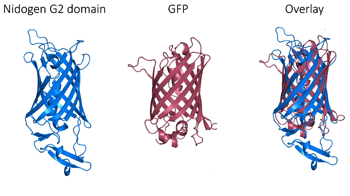
Despite these very promising results, GFP is an exogenous protein from Aequorea victoria and, consequently, triggers an immune response, which limits its clinical use. Thus, we needed to find a human protein that matches the exceptional properties of GFP as a protein scaffold. Fortunately, a non-fluorescent GFP-like protein has been described in humans and it corresponds to one of the three globular domains of Nidogen, a structural protein that binds to collagen IV, laminin and perlecan with high affinity. The globular domain G2 has a beta-barrel structure with a central alpha-helix that folds very similarly to the GFP, despite that these proteins share very low sequence identity. Notably, this domain does not have post-translational modifications that could interfere with its production and folding in prokaryotic cells.

However, perlecan and collagen IV binding sites have been reported within this G2 domain. Therefore, we needed to selectively mutate these binding sites in order to assure the biological neutrality of the nanoparticles. After a thorough structural analysis, we incorporated four different mutations to engineer a biologically neutral product that was named HSNBT. There were no differences detected between the wild-type G2 domain and the engineered HSNBT protein regarding the predicted structural epitopes, which suggested that the introduced mutations would not generate immunogenicity.

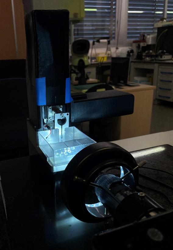
In order to validate the new scaffold, we used the above-mentioned patented platform with T22 and the hexa-histidine tag, replacing GFP for the new HSNBT scaffold. First, we characterized the resulting nanoparticles and we determined, both by Dynamic Light Scattering (DLS) and Scanning Electron Microscopy (SEM), that they had a size of around 10 nanometers. Then, we observed that the T22-HSNBT-H6 nanoparticles were internalized effectively by CXCR4+ cells. This specificity was corroborated when we used a CXCR4 antagonist (AMD) and we saw a notable decrease of their internalization. Then, we successfully conjugated floxuridine to the nanoparticles (T22-HSNBT-H6-FdU) through the free lysine-amino groups of the protein and we demonstrated that the nanoconjugates had a potent cytotoxic effect in CXCR4+ cells.

Once we have validated these nanoconjugates in vitro, we tested them in a colorectal cancer mouse model. Notably, we saw an important tumor growth inhibition after several doses of these nanoconjugates. The inhibitory effect was slightly higher when using the new scaffold than with GFP. We also saw a significant increase in cell death bodies and caspase-3 activation in the tumor after the treatment with the nanoconjugates. Again, the effect was more potent with HSNBT as a scaffold than with GFP. Remarkably, the treatment did not result in any histological toxicity and there were no differences between the weight of the treated mice when compared to the untreated mice.

This technology is protected by 3 patents: The ligand to enter CXCR4+ cells (WO2012/095527), the nanoconjugates (EP17382461.6) and the human scaffold protein HSNBT, (EP19383201), all three licensed to Nanoligent SL.

All in all, these results confirm that the G2 domain of nidogen can be used as a protein scaffold for targeted drug delivery. Its performance both in vitro and in vivo not only matches the observed with GFP, but it is even more efficient than GFP when conjugated with floxuridine. Therefore, the engineered HSNBT protein shows a very exciting potential to be used in the development of protein-based nanomedicines.  

By Carlos Martínez Torró (NANBIOSIS U1 PPP)

Read More

U16-S07. Evaluation of the adhesion capacities and formation of biolayers of microorganisms on materials. Evaluation of bactericidal and/or bacteriostatic responses of materials. OUTSTANDING

Description:

Evaluation of the adhesion of microorganisms to surfaces, including the evaluation of their viability. Adhesion can be studied under flow or static and at different contact times. Study of biolayers formed on materials after different growth times. Evaluation of bactericidal and/or bacteriostatic responses using viability kits or plate growth.


Applications:

Testing of materials with anti-infective objectives. Testing of compounds with antibacterial purpose.

Read More

U16-E07. Equipment for microbiology tests on materials. OUTSTANDING

Description: It is the set of precise elements to carry out microbiological tests with materials. Includes laminar flow adhesion chambers, Robbin devices, shaker and drip bioreactors, fluorescence microscopes, plate readers, ovens, autoclaves, etc.

Technical specifications: Allows quantifications on different types of systems. Depending on the specific conditions, it is possible to evaluate behavior in extensive solid materials or in suspension.

Applications: The services that can be provided with this equipment are:
Evaluation of the adhesion capacities and formation of biolayers of microorganisms on materials. Evaluation of bactericidal and/or bacteriostatic responses of materials.

Read More

U16-S06. Topographic characterization service using optical profilometry OUTSTANDING

This service makes it possible to determine the surface tension of both solids and liquids under controlled temperature and humidity conditions, even locally by means of deposited drops with a volume of around 10-12 L. The service will be provided by specialists in the characterization of materials in dimensions macroscopic or in colloidal systems. The service can meet the needs of business R&D&i, providing support for their controls according to ISO, ASTM, ASME and EUR standards. In addition, it can also serve research groups with detailed reports according to the needs of the study.

Read More

U16-S05. Characterization service by contact angle goniometry OUTSTANDING

This service makes it possible to determine the surface tension of both solids and liquids under controlled temperature and humidity conditions, even locally by means of deposited drops with a volume of around 10-12 L. The service will be provided by specialists in the characterization of materials in dimensions macroscopic or in colloidal systems. The service can meet the needs of business R&D&i, providing support for their controls according to ISO, ASTM, ASME and EUR standards. In addition, it can also serve research groups with detailed reports according to the needs of the study.

Read More

1st Forum of CIBER-BBN/NANBIOSIS and CSIC Nanomed Conection researchers

The Nanomed Conection of the Spanish Research Council (CSIC) and the Networking Biomedical Research Center in Bioengineering, Biomaterials and Nanomedicine (CIBER-BBN), together with its singular infrastructure NANBIOSIS, have organised a Forum on Nanomedicine Research during the days 30 of June and 1st of July to be held at the Institute of Advanced Chemistry of Catalonia (IQAC-CSIC) in Barcelona. The event will be also transmitted on-line previous registration.

This is the first meeting gathering together scientists from CIBER-BBN and its ICTS NANBIOSIS and from the CSIC’ Nanomed Conection with a shared interest in Nanomedicine.

This two days meeting will allow researchers to present their works in progress, share their scientific concerns and needs and discuss the impact of nanomedicine in the emerging fields of drug delivery, diagnosis and therapy.

The programe, available in the web of the forum includes these sessions:

  • Nanobiotechnological solutions for diagnosis and therapy
  • Drug delivery nanosystems
  • Applications for oncology (I and II)
  • Nanomedicine & other frontier applications

Attendance to the Forum (in person / or online) is free prior registration in the web of the forum (following this link):

Registration will remain open until June 26.
We hope to see you there!

Read More

NANBIOSIS in the new poster of ICTS map published by Spanish Goverment.

ICTS underpins the Spanish reputation for research excellence.

In the picture: the new poster of the ICTS map in which NANBIOSIS facilities have been highlighed

The term Unique Scientific and Technical Infrastructure (ICTS) refers to facilities, resources, or services for the development of top-quality cutting-edge research, as well as the communication, exchange, and preservation of knowledge, the transfer of technology, and promotion of innovation. They are unique or exceptional in their fields, with a high cost of investment, maintenance, and operation, and are of a strategic importance that justifies their availability to all actors in the field of R&D&I. The ICTS share three fundamental characteristics; they are infrastructures with public ownership, unique and open to competitive access.

ICTS offer an opening capacity percentage of their essential services under ‘Competitive Open Access’ for the use by national and international public and private sector researchers, with the support of technical and administrative personnel of the ICTS. Infrastructures access is ruled by a public “Access Protocol” that describes the procedure and criteria for access to the infrastructure. The main features of ‘Competitive Open Access’ are that R + D + i quality of activities developed at the infrastructure should be proven and that requests for access should be prioritized on the basis of objective criteria.

The dissemination of ICTS and their capabilities is essential to provide Spanish and international researchers with access to a large base of quality services and facilities, a basic requirement for the development of excellent science.

The new posster pushised by the General Subdirectorate of Large
Scientific-Technical Facilities of the of the Sapanish Ministry of Science and Research Innovation helps to disseminate and understand the map of ICTS wich are located throughout the country

NANBIOSIS, is one of the five ICTS in the field of Health Sciences and Biotecnology

Read More

U13-E04. Micro-scale tension-compression test system CellScale.

Test system for small samples including:

  • Main equipment (Micro-scale tension-compression test system)
  • Tangential test module (Shear Axis add-on for MicroTester system)

Description: Enhanced for smaller samples, the MicroTester offers improved force resolution, streamlined test setups, and exceptional visual feedback. It’s ideal for a wide range of applications, from tiny tissue samples to hydrogel microspheres, cell spheroids, and engineered microtissues.

Technical specifications: Piezo-electric actuators with 0.1µm resolution Optional second axis imaging Force resolution down to 10nN High resolution CCD imaging Integrated temperature-controlled media bath Fully featured user interface software for simple, cyclic, relaxation, and multi-modal testing with real-time feedback

Aplications: Compression, tension, bending, indentation and shear testing

Read More

U13-S09. COMPRESSION-SHEAR TEST CELLSCALE MicroTester G2 (On-site) OUTSTANDING

U13-S09. COMPRESSION-SHEAR TEST CELLSCALE MicroTester G2 (On-site) OUTSTANDING

Uniaxial tensile and shear tests

Description: By detecting small changes in a force sensing micro-wire, the system can measure forces and displacements that are much smaller than a conventional mechanical test system. The system incorporates a temperature-controlled fluid bath for maintaining ideal specimen conditions and a high-quality optical system to provide user feedback

Aplications: Tension and compression and shear test of hydrogel microspheres, cell spheroids, and engineered microtissues

Read More

How to accomplish researchers’ goals with Confocal Microscopy: the tools, the know-how and the expertise you need

NANBIOSIS Unit 17 (Confocal Microscopy) is a CIBER-BBN unit located in the Cell Culture Unit, CAI Medicine and Biology, Faculty of Medicine at the University of Alcala. This unit of the ICTS NANBIOSIS supports researchers interested on their different studies visualizing diverse samples as tissues, cells, bacterial biofilms, etc. This unit owns the tools, the know-how and the expertise to accomplish researchers’ goals either by transmission or reflection fluorescent.

We are happy of sharing this video in which researchers of Unit 17 show all the steps required for the visualization of the PV-1 molecule, also known as PLVAP, on the gut-endothelium of cirrhotic rats. We look at the whole process, starting by the sample selection following their preparation until its visualization by the confocal fluorescent microscopy, ending up with the analyze process.

Read More