+34 620 10 75 37info@nanbiosis.com

Posts on Jan 1970

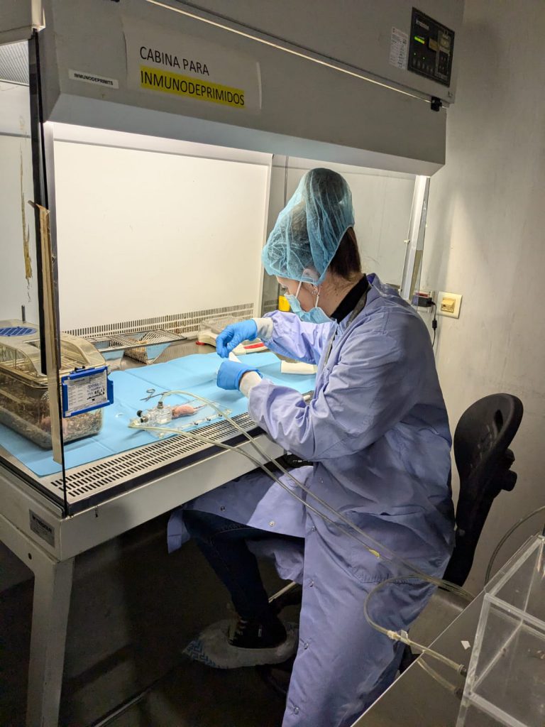
U22-E01. Rooms for the maintenance of large animals

This unit has facilities with a perimeter area of 3,200 m2 and it has large rooms to house the experimental animals according to their species characteristics. Among the equipment and facilities include:
9 rooms for large animals (pigs) maintenance with capacity for about 350 animals, for long periods of time and environmental, nutritional and microbiological conditions controlled.

Read More

U22-E02. Rooms for maintaining small ruminants

2 rooms for the maintenance of small ruminants (sheep and goats) with capacity for about 60 animals over long periods of time and environmental, nutritional and microbiological conditions controlled.

Read More

U22-S01. Animal Housing (Remote) OUTSTANDING

It houses different animal species, whether small or big (rats, mice, rabbits, dogs, pigs, sheep, etc), in compliance with the physical and legal conditions for experimental research.

Read More

U18-S01. In vitro Toxicology

In vitro Toxicology

The aim of this service is to evaluate the in vitro toxicity of compounds in different types of normal human cells in which we determine the effect using different types of assays including MTT, LDH release and caspase activation. The cellular models that we offer include cells from kidney, liver, bone marrow stroma or peripheral blood. Additionally, we are offering immunology assays including detection of NO production and chemotaxis and phagocytosis assays.

Customer benefits

The costumer will benefit from more than 20 years of experience of the researchers involved in the service that have performed the in vitro evaluation of the toxicity of different nanoparticles and other types of compounds. We offer also the possibility of setting up new in vitro assays or to offer other cellular models that could be of interest for the costumer. Moreover, the conditions of the assays are flexible and will be adapted to the needs of each specific compound.

Target customer

The offered service can be of interest to research groups of academia or companies willing to test the toxicity of any compound in vitro.

References

  • Falgàs A, Pallarès V, Unzueta U, Núñez Y, Sierra J, Gallardo A, Alba-Castellón L, Mangues MA, Álamo P, Villaverde A, Vázquez E, Mangues R, Casanova I. Specific Cytotoxic Effect of an Auristatin Nanoconjugate Towards CXCR4+ Diffuse Large B-Cell Lymphoma Cells. Int J Nanomedicine. 2021 Mar 5;16:1869-1888. doi: 10.2147/IJN.S289733.
  • Núñez Y, Garcia-León A, Falgàs A, Serna N, Sánchez-García L, Garrido A, Sierra J, Gallardo A, Unzueta U, Vázquez E, Villaverde A, Mangues R, Casanova I. T22-PE24-H6 Nanotoxin Selectively Kills CXCR4-High Expressing AML Patient Cells In Vitro and Potently Blocks Dissemination In Vivo. Pharmaceutics. 2023 Feb 22;15(3):727. doi: 10.3390/pharmaceutics15030727.
Read More

U18-S02. In vivo Nanotoxicology

In vivo Nanotoxicology

The aim of this service is to evaluate the in vivo biodistribution, toxicity and therapeutic index of nanoparticles and drug-loaded nanoparticles. Thus, we offer different mouse models in which we can determine the biodistribution of nanoparticles, with or without loaded drug, in normal and tumor tissues. Moreover, we can also determine the toxicity of the compounds and their antitumor activity in different tumor models. In the service we have subcutaneous and orthotopic/disseminated models of lymphoma, leukemia and colorectal, endometrium, head and neck carcinomas. Moreover, we have also available PDX models of colorectal and endometrium cancer. We can also offer the possibility to set up new animal models of other tumor types.

Customer benefits

The costumer will benefit from the experience of the researchers involved in the service that have performed the in vivo evaluation of a high number of diverse nanoparticles. The service has also high expertise in developing new cancer animal models. Thus, we offer also the possibility of setting up new models of any cancer type that fit with the needs of the costumer. Moreover, the conditions of the assays are flexible and will be adapted to the need of each specific compound.

Target customer

The offered service can be of interest to research groups of academia or companies willing to test the biodistribution, toxicity or antitumor activity of nanoparticles or drug-loaded nanoparticles in vivo.

References

  • Martínez-Torró C, Alba-Castellón L, Carrasco-Díaz LM, Serna N, Imedio L, Gallardo A, Casanova I, Unzueta U, Vázquez E, Mangues R, Villaverde A. Lymphocyte infiltration and antitumoral effect promoted by cytotoxic inflammatory proteins formulated as self-assembling, protein-only nanoparticles. Biomed Pharmacother. 2023 Aug;164:114976. doi: 10.1016/j.biopha.2023.114976.
  • Rioja-Blanco E, Arroyo-Solera I, Álamo P, Casanova I, Gallardo A, Unzueta U, Serna N, Sánchez-García L, Quer M, Villaverde A, Vázquez E, Mangues R, Alba-Castellón L, León X. Self-assembling protein nanocarrier for selective delivery of cytotoxic polypeptides to CXCR4+ head and neck squamous cell carcinoma tumors. Acta Pharm Sin B. 2022 May;12(5):2578-2591. doi: 10.1016/j.apsb.2021.09.030.
  • Falgàs A, Garcia-León A, Núñez Y, Serna N, Sánchez-Garcia L, Unzueta U, Voltà-Durán E, Aragó M, Álamo P, Alba-Castellón L, Sierra J, Gallardo A, Villaverde A, Vázquez E, Mangues R, Casanova I. A diphtheria toxin-based nanoparticle achieves specific cytotoxic effect on CXCR4+ lymphoma cells without toxicity in immunocompromised and immunocompetent mice. Biomed Pharmacother. 2022 Jun;150:112940. doi: 10.1016/j.biopha.2022.112940.
    -Medina-Gutiérrez E, García-León A, Gallardo A, Álamo P, Alba-Castellón L, Unzueta U, Villaverde A, Vázquez E, Casanova I, Mangues R. Potent Anticancer Activity of CXCR4-Targeted Nanostructured Toxins in Aggressive Endometrial Cancer Models. Cancers (Basel). 2022 Dec 23;15(1):85. doi: 10.3390/cancers15010085.
  • Sala R, Rioja-Blanco E, Serna N, Sánchez-García L, Álamo P, Alba-Castellón L, Casanova I, López-Pousa A, Unzueta U, Céspedes MV, Vázquez E, Villaverde A, Mangues R. GSDMD-dependent pyroptotic induction by a multivalent CXCR4-targeted nanotoxin blocks colorectal cancer metastases. Drug Deliv. 2022 Dec;29(1):1384-1397. doi: 10.1080/10717544.2022.2069302.
  • Serna N, Falgàs A, García-León A, Unzueta U, Núñez Y, Sánchez-Chardi A, Martínez-Torró C, Mangues R, Vazquez E, Casanova I, Villaverde A. Time-Prolonged Release of Tumor-Targeted Protein-MMAE Nanoconjugates from Implantable Hybrid Materials. Pharmaceutics. 2022 Jan 14;14(1):192. doi: 10.3390/pharmaceutics14010192.
  • Pallarès V, Unzueta U, Falgàs A, Aviñó A, Núñez Y, García-León A, Sánchez-García L, Serna N, Gallardo A, Alba-Castellón L, Álamo P, Sierra J, Cedó L, Eritja R, Villaverde A, Vázquez E, Casanova I, Mangues R. A multivalent Ara-C-prodrug nanoconjugate achieves selective ablation of leukemic cells in an acute myeloid leukemia mouse model. Biomaterials. 2022 Jan;280:121258. doi: 10.1016/j.biomaterials.2021.121258.
Read More

U17-E01. Leica TCS-SP5 confocal microscope

Leica TCS-SP5 confocal microscope with especial features that allows studying interactions between cells/tissues and materials. Indeed, the experience of the research group in charge of this Unit makes this a unique service for the study of cells and tissues and the interactions between various materials and cell components as well as between implants/scaffolds and tissues of the recipient organism. For example, it makes it possible to characterize in detail the specific markers of particular cell populations for tissue engineering applications and in vitro tests of biocompatibility of new materials, allowing the process of cell colonization of surfaces to be explored and simultaneous analysis of the cells or subcellular structures and materials. The combined use of fluorescence and reflection makes it possible to study the tissue-implant interface, with up to 8 fluorophores, at wavelengths from 405 to 633 nm allowing histological studies of a wide range of tissue types which would be restricted at certain fixed wavelengths due to autofluorescence phenomena.


Leica TCS-SP5 confocal microscope with:

DMI 6000 inverted microscope with 4 objectives:
10X (dry); 20X; 40X and 63X(for immersion).

Confocal module:
•• 3 channels for spectral detection
•• AOBS (Acousto Optical Beam Splitter)
•• Resonant scanning system.

4 lasers:
•• Argon laser (excitation at 458, 477, 488, 496 and 514 nm)
•• He/Ne laser (excitation at 633 nm)
•• DPSS (diode-pumped solid-state) laser (excitation at 561 nm)
•• UV laser (excitation at 405 nm)

Incubation chamber on a motorized stage.

Workstation with 4 types of software for data acquisition and processing:

•• 3D imaging
•• Colocalization analysis
•• FRAP (Fluorescence Recovery After Photobleaching)
•• FLIP (Fluorescene Loss In Photobleaching)
•• FRET (Fluorescence Resonance Energy Transfer)

Read More

U17-S01. Confocal Microscopy Service

Confocal Microscopy Service

The Confocal Microscopy Service offers the possibility of making optical section of samples and enabling their three-dimensional reconstruction, which allows their visualization in high resolution with exceptional clarity and detail.
The unit is equipped with a Leica TCS-SP5 confocal microscope. The confocal module is equipped with three spectral detection channels, AOBS (Acousto-optical beam splitter) and a resonant scanner system that allows analysis at high speed and resolution and makes possible the analysis of dynamic in vivo physiological processes in short periods of time, significantly improving the survival of living biological samples by shortening the exposure times to lasers. Includes 1 argon laser, 1 He/Ne laser, 1 DPSS laser diode and 1 violet excitation laser. The microscope is coupled to a cell incubation kit that allows multi-position time lapse experiments. The equipment includes a workstation and four software for acquisition and analysis, which allow 3D visualizations, co-location studies, FRAP (Fluorescent Recovery after Photo-bleaching), FLIP (Fluorescent Loss in Photobleaching) and FRET (Fluorescence Resonant Energy Transfer). The equipment allows 3D characterization in detail of living cells and tissues through the use of different fluorochromes, expression and localization of molecules in 2/3D, colocalization and interaction of proteins or other types of molecules; endocytosis and intracellular transport, in situ hybridization with fluorescent probes, interaction studies between cells and materials, etc.
The unit provides researchers with a wide array of routine and specialized services as well as the latest advances in microscopy, including technical and scientific support to scientists for the study of cell/tissue biology, physiology and pathogenesis of diseases. Overall, the Confocal Microscopy Service serves as a valuable tool to advance scientific research and understand complex biological systems at the cellular and molecular level.

Customer benefits

Confocal microscopy is useful in biomedical science, cell and molecular biology and physiology for visualizing subcellular organelles, cellular interactions, disease mechanisms, co-localization studies, analysis of gene expression or in vivo and real-time experiments using markers or fluorescent fusion proteins.
Also, in tissue engineering and materials science, confocal microscopy enables surface morphology characterization, and material properties.

Confocal Microscopy Service offers specific advantages to customers by providing high-resolution imaging and adding value through access to advanced instrumentation and expertise. It becomes essential for organizations involved in Research and Development across various sectors, enabling them to advance scientific understanding, accelerate innovation, and drive discoveries that have social impact.

Target customer

The Confocal Microscopy Service becomes essential for organizations involved in Research and Development in various scenarios:

  • Biomedical Research Institutes: This service can be useful for research institutions focused on the study of cellular and molecular mechanisms underlying different diseases, allowing to visualize pathological changes, identify biomarkers and investigate possible therapeutic targets. Additionally, it allows researchers to study disease progression, evaluate drug efficacy, and develop new treatment strategies.
  • Pharmaceutical Companies: Confocal microscopy can be used by drug development companies to evaluate the effects of these drugs on cellular function.
  • Academic Laboratories: Confocal microscopy can be useful for researchers in diverse fields of biology, including neuroscience, developmental biology, and cancer biology, to explore fundamental biological processes and generate novel scientific insights.

References

  1. Benito-Martínez S, Pérez-Köhler B, Rodríguez M, García-Moreno F, Gómez-Gil V, Pascual G, Bellón JM. Antibacterial Biopolymer Gel Coating on Meshes Used for Abdominal Hernia Repair Promotes Effective Wound Repair in the Presence of Infection. Polymers (Basel). 2021;13(14):2371. doi: 10.3390/polym13142371.
  2. Sánchez-Esteban S, Castro-Pinto M, Cook-Calvete A, Reventún P, Delgado-Marín M, Benito-Manzanaro L, Hernandez I, López-Menendez J, Zamorano JL, Zaragoza C, Saura M. Integrin-Linked Kinase Expression in Human Valve Endothelial Cells Plays a Protective Role in Calcific Aortic Valve Disease. Antioxidants (Basel). 2022. 11(9):1736. doi: 10.3390/antiox11091736. 
  3. Campillo S, Bohorquez L, Gutiérrez-Calabrés E, García-Ayuso D, Miguel V, Griera M, Calle Y, de Frutos S, Rodríguez-Puyol M, Rodríguez-Puyol D, Calleros L. Indoxyl sulfate- and P-cresol-induced monocyte adhesion and migration is mediated by integrin-linked kinase-dependent podosome formation. Exp Mol Med. 2022;54(3):226-238. doi: 10.1038/s12276-022-00738-8. 
Read More

U16-E04. ToF-SIMS 5 Time-of-flight secondary-ion mass spectrometry system (ION TOF )

ToF-SIMS 5 Time-of-flight secondary-ion mass spectrometry system (ION TOF ).

•• Sensitivity < 100 ppm.
•• Lateral resolution ~ 100 nm (extension of surface area analyzed).
•• Depth resolution ~ 1 nm (thickness of surface analyzed).
•• Identification of molecules up to 10000 amu.

Read More

U16-E03. K-Alpha X-ray photoelectron spectroscopy (XPS) system (Thermo)

K-Alpha X-ray photoelectron spectroscopy (XPS) system (Thermo):

•• Lateral resolution ~ 30-400 μm (extension of surface area analyzed).
•• Depth resolution ~ 10 nm (thickness of surface analyzed).
•• Depth profiles.

Read More

U16-E02. UVISEL ellipsometer (Horiba JovinYvon)

UVISEL ellipsometer (Horiba JovinYvon), range 190-2100 nm, based on phase modulation with cell for liquid media:

•• Measurement of thicknesses from 0.1 nm to 30 μm.
•• Measurement of thickness of various superimposed layers.
•• Simultaneous measurement of the proportion of various different materials present on a surface.
•• Estimation of the porosity as percentage of pores vs. deposited material.
•• Measurement of roughness of coating between 2.5 and 15 nm.

Read More