+34 620 10 75 37info@nanbiosis.com

Posts on Jan 1970

U12-E18. Viscosimeter RVDV-II+P (Brookfield)

U12-E18. Viscosimeter RVDV-II P (Brookfield)

Description:
Brookfield viscometer with a wide measurement range from 25 mPa.s to 64 MmPa.s. It is equipped with an accessory (Small Sample Adaptor SSA) for samples of small volume (7-13.5 mL).

Applications:
Viscosity measurements.

U12-E18. Viscosimeter RVDV-II P (Brookfield)
Read More

U12-E17. Zetasizer NanoZ (Malvern Instruments)

U12-E17. Zetasizer NanoZ (Malvern Instruments)

Description:
Instrument for the measurement of zeta potential, using the M3-PALS technology for the determination of electrophoretic mobility.  Temperature can be controled from 2 to 90 ˚C. It uses disposable capillary cells “Universal Dip Cell” for standard Zeta potential measurements in organic and aqueous solvents, and it can use the “High Concentration Cell“ for measurements at high concentrations

Applications:
Measurement of electrophoretic mobility and zeta potential in dispersions

U12-E17. Zetasizer NanoZ (Malvern Instruments)
Read More

U12-E16. Autosorb IQ (Quantachrome Instruments)

U12-E16. Autosorb IQ (Quantachrome Instruments)

Description:
The Quantachrome Autosorb iQ-AG equipment determines gas adsorption/desorption curves, for the calculation of specific surface area and pore size analysis. This equipment allows the simultaneous degassing of two samples and the physisorption analysis of a third sample, at relative pressures above P/P0 > 1×10⁻³. The gas sorption analyzer is equipped with a cryogenic dewar for measurements until 90 hours.

Applications:
Characterization of the porosity and surface area of nanoparticles, macroporous, macro/mesoporous and mesoporous materials like aluminas, silicas and other materials including M41S materials (eg. MCM-41), PMOs, KITs and many SBAs.

.

U12-E16. Autosorb IQ (Quantachrome Instruments)
Read More

U12-E15. SEM Hitachi TM-1000 (Hitachi)

U12-E15. SEM Hitachi TM-1000 (Hitachi)

Description:
Tabletop Scanning Electron Microscope.

Applications:
Observations of the morphology of solid samples, such as polymeric and synthetic particles, meso/macroporous materials, textiles, etc., with high field depth (it allows focusing simultaneously on a wide portion of the sample) and relatively high resolution (in the nanometer range). Samples do not need to be electrical conductive, and therefore, coating with a very thin metal layer (e.g. Au, Pd) is not always required. The only sample requirement is the absence of volatile components, including water humidity

U12-E15. SEM Hitachi TM-1000 (Hitachi)
Read More

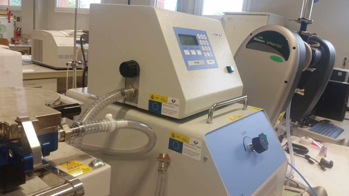
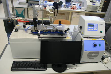
U10-E38 Equipment of biopharmaceutical characterization of pulmonary formulations with Breath Simulator and Alberta Idealised Throat (AIT)

Equipment of biopharmaceutical characterization of pulmonary formulations with Breath Simulator and Alberta Idealised Throat (AIT)


1) NGI Cascade Impactor: It’s an analytical tool for the development of inhalation products. It’s use for the testing of all inhalation formulations and devices: MDIs, DPIs, nebulizers and aerosol and nasal sprays. It’s the new “workhorse” of the pharmaceutical industry.

2) Alberta Idealised Throat (AIT): before entering the lungs, aerosols must traverse the mouth-throat. So it’s important to be able to mimic aerosol deposition and flow in the human mouth-throat when studying inhaled aerosols. The Alberta geometry has been shown to do a remarkable job of mimicking the aerosol and flow motion in the human mouth-throat and allow researchers examining  and testing aerosols inhaled orally.

3) Breath Simulator: are increasingly used in testing orally inhaled and nasal drug products (OINDPs) to replace existing constant flow conditions with breathing profiles more representative of conditions in vivo. It can be used for improved IVIVC applications, with real breath profiles generated in clinic.

Read More

U10-E37. 3D Bioprinter

3D Bioprinting

Read More

U10-E35 Confocal Microscopy Unit

Confocal Microscopy Unit

Read More

JUMISC, partner of NANBIOSIS, represents Spain in a European network of 22 countries

The Jesús Usón Minimally Invasive Surgery Centre (JUMISC), partner of NANBIOSIS toguether with CIBER-BBN,  leads a multidisciplinary European research network to improve present urinary stents and prevent their causes of failure.

The idea of creating the network was born from a research, started in 1999 by Doctor Soria, Researcher-Coordinator of the Endoscopic Surgery Area at the JUMISC and the promoter of the mentioned network, representing Spain.

His proposal has been supported by the European and Spanish Urology Association, and funded by the Horizon 2020 Program-Cost Actions (European Cooperation in Science and Technology)

The focus of this Action is to create a multidisciplinary team to identify the problems concerning the urinary stents (design, composition, biomaterial applications, coatings, incrustation, etc.) and to promote future research on this field. In this way, the network will try to reduce adverse effects provoked by the present stents.

This European network (2017-2021), coordinated by Dr. Soria, the Chair of the Action, is composed by 22 European countries and other proposals coming from USA,  Japan, South Korea,  Israel, Canada, Russia, India and it is open to other invited countries to cooperate inside  the organised working groups.

This action will provide, during the next 4 years, an extensive and interdisciplinary R&D program to discover the causes of failure of the present stents and will propose cooperative work in urology and bioengineering field.

This proposal, an initiative of the JUMISC, leaded by Doctor Sánchez Margallo, will contribute to improve healthy-quality of patients, reduce in health care costs, and increase the competitiveness of the European medical device industry.

Read More

New Zealand White rabbit as a model for preclinical studies addressing tissue repair at the level of the abdominal wall.

Juan Manuel Bellón and Gemma Pascual, Scientific Director and Coordinator of Unit 17 of NANBIOSIS, have recently published an article in the prestigious journal Tissue Eng Part C Methods.

Once a prosthetic material is implanted, an inflammatory reaction is induced in an attempt to remove it or isolate it from the host tissue. After this initial response, a cellular response takes place that gives rise to the start of the process of integration of the biomaterial within the host tissue. Immunohistochemical studies can provide information on the events that regulate this process, and are useful to analyze the progression of the implant and the neoformed tissue. For the tissue repair of a damaged area constantly subjected to tensile forces such as the abdominal wall, optimal collagenization is an extremely important factor. The proper deposition and organization of mainly collagen type I in the neoformed scar tissue will condition the mechanical strength and compliance of the repair zone and thus possible hernia recurrence. In this report, we review the use of the New Zealand White rabbit as the experimental animal for several models of abdominal wall repair and among other techniques, collagen component of the implants was assessed by immunofluorescence using anti-collagen type I and type III monoclonal antibodies. Samples were examined using a confocal microscope Leica SP5 belonging to the platform ICTS ‘NANBIOSIS’ U17 of the CIBER-BBN at the University of Alcalá.

Important information gained from selected models was observed in terms of tissue ingrowth and collagen deposition in the implant area showing significant differences depending on the properties of the implanted prosthetic material. Some variations were also observed in biomechanical properties according to the components comprising each prosthetic material.

Read More