+34 620 10 75 37info@nanbiosis.com

Blog

Fabry Desease in the Rare Disease Day: A New Hope

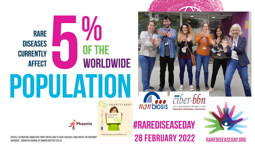
WHY DO CELEBRATE TODAY THE INTERNATIONAL #RareDiseaseDay?

29 of February is a ‘rare’ date and February, a month with a ‘rare’ number of days, has become a month to raise awareness about rare diseases and their impact on patients’ lives.  Since 2008 thousands of events happen every year all around the world and around the last day of February with the aim of improving equity and reducing stigmatization for people who live with more than 6,000 rare diseases.

WHAT ARE RARE DISEASES

Rare diseases are pathologies or disorders that affect a small part of the population (less than 5 per 10,000 inhabitants) and generally have a genetic component. They are also known as orphan diseases.

Diseases present a series of particular symptoms, and it is very difficult to diagnose what their true cause is. These disorders or alterations that patients present must be evaluated by a specialist, depending on each case.

Today 5% of the world population suffer from them. This translated into numbers, corresponds to approximately 300 million affected.

A patient with a rare disease waits an average of 4 years to obtain a diagnosis, in 20% of cases it takes 10 or more years to achieve the proper diagnosis.

ORPHAN DRUGS

To combat this disease, patients need to be treated with so-called orphan drugs. They serve to prevent and treat pathology. Its composition is based on biotechnological compounds whose manufacture is very expensive and not profitable for companies. For this reason, cooperation of governments is needed as well as financial incentives to encourage pharmaceutical companies to develop and market medicines to make these treatments accessible to a greater number of people.

FABRY DISEASE

Fabry is one of the rare diseases that currently lack a definitive cure. Symptoms may include episodes of pain, especially in the hands and feet (acroparesthesias); small dark red spots on the skin called angiokeratomas; decreased secretion of sweat (hypohidrosis); opacity of the cornea (cataracts) and hearing loss. Internal organs such as the kidney, heart, or brain may be involved, resulting in progressive kidney damage, heart attacks, and strokes.

Fabry disease is a lysosomal storage disease arising from a deficiency of the enzyme α-galactosidase A (GLA). The enzyme deficiency results in an accumulation of glycolipids, which over time, leads to cardiovascular, cerebrovascular, and renal disease, ultimately leading to death in the fourth or fifth decade of life. Currently, lysosomal storage disorders are treated by enzyme replacement therapy (ERT) through the direct administration of the missing enzyme to the patients.

SMART 4 FABRY” EUROPEAN PROJECT

CIBER-BBN, through the researcher Nora Ventosa has coordinated the european project “Smart-4-Fabry” developed during 2017-2021, the proyect was undertaken by a consortium formed by ten partners, including private companies and public institutions in Europe and Israel, with a Horizon 2020 financial programme by the European Commission (H2020-NMBP-2016-2017; call for nanotechnologies, advanced materials, biotechnology and production; Proposal number: 720942-2).

In view of their advantages as drug delivery systems, liposomes are increasingly being researched and utilized in the pharmaceutical, food and cosmetic industries, but one of the main barriers to market is their scalability.

Depressurization of an Expanded Liquid Organic Solution into aqueous solution (DELOS-susp) is a compressed fluid-based method that allows the reproducible and scalable production of nanovesicular systems with remarkable physicochemical characteristics, in terms of homogeneity, morphology, and particle size. The objective of this work was to optimize and reach a suitable formulation for in vivo preclinical studies by implementing a Quality by Design (QbD) approach, a methodology recommended by the FDA and the EMA to develop robust drug manufacturing and control methods, to the preparation of α-galactosidase-loaded nanoliposomes (nanoGLA) for the treatment of Fabry disease.

Through a risk analysis and a Design of Experiments (DoE), researechers obtained the Design Space in which GLA concentration and lipid concentration were found as critical parameters for achieving a stable nanoformulation. This Design Space allowed the optimization of the process to produce a nanoformulation suitable for in vivo preclinical testing.

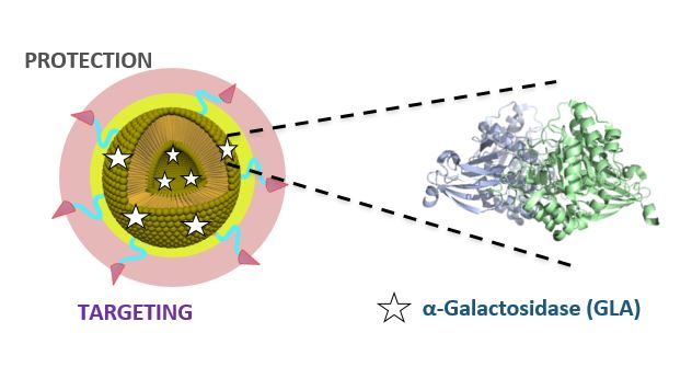
The new nanoformulation developed by Smart4Fabry for the treatment of Fabry disease achieved the ODD (Orphan Drug Designation) by the European Commission. The new nanomedicine is more effective and has a better biodistribution than the current treatments, based on enzyme replacement. The new nanomedicine is based on a nanovesicle that protects the enzyme and achieves a better cell internalisation, thus reducing the doses needed, the total cost and improving the quality of patients.

Four units of NANBIOSIS participated in the project:

– U1 Protein Production Platform (PPP) led by Neus Ferrer and Antony Villaverde at IBB-UAB for the production and purification in different expression systems for R&D purposes.

– U3 Synthesis of Peptides Unit led by Miriam Royo at IQAC-CSIC performed all the chemical process of the Smart-4-Fabry project, i.e. design and synthesis of peptides used as targeting ligands in the nanoliposome formulation.

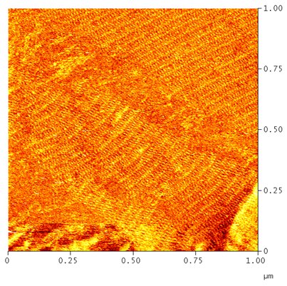
– U6 Biomaterial Processing and Nanostructuring Unit led by Nora Ventosa at ICMAB-CSIC developed tasks related to the manufacture of the nanoliposome formulation of GLA enzyme and the physico-chemical characterization (this unit counts with plants at different scales, from mL to L, which allow process development by QbD and process scale-up, as well as instrumental techniques for assessment of particle size distribution, particle concentration, particle morphology and stability, and Z-potential) .

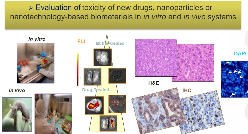
– U20 In Vivo Experimental Platform led by Ibane Abásolo at VHIR carried out the non-GLP preclinical assays of the project (in vivo efficacy, biodistribution and tolerance/toxicity assays).

PHOENIX: OPEN INNOVATION TEST BED

Researchers of CIBER-BBN and NANBIOSIS, led by Nora Ventosa, are currently participating in another european project, PHOENIX “Enabling Nano-pharmaceutical Innovative Products” in the framework of which this novel nanomedicine developed under the Smar4Fabry project and designed as Orphan Drug by the EMA, will be scaled-up and manufactured under GMP to enable its clinical testing.

Articles of reference:

Josep Merlo-Mas, Judit Tomsen-Melero, José-Luis Corchero, Elisabet González-Mira, Albert Font, Jannik N. Pedersen, Natalia García-Aranda, Edgar Cristóbal-Lecina, Marta Alcaina-Hernando, Rosa Mendoza, Elena Garcia-Fruitós, Teresa Lizarraga, Susanne Resch, Christa Schimpel, Andreas Falk, Daniel Pulido, Miriam Royo, Simó Schwartz, Ibane Abasolo, Jan Skov Pedersen, Dganit Danino, Andreu Soldevila, Jaume Veciana, Santi Sala, Nora Ventosa, Alba Córdoba, “Application of Quality by Design to the robust preparation of a liposomal GLA formulation by DELOS-susp method”, The Journal of Supercritical Fluids, Volume 173, 2021, 105204, https://doi.org/10.1016/j.supflu.2021.105204.

Judit Tomsen-Melero, Solène Passemard, Natalia García-Aranda, Zamira Vanessa Díaz-Riascos, Ramon González-Rioja, Jannik Nedergaard Pedersen, Jeppe Lyngsø, Josep Merlo-Mas, Edgar Cristóbal-Lecina, José Luis Corchero, Daniel Pulido, Patricia Cámara-Sánchez, Irina Portnaya, Inbal Ionita, Simó Schwartz, Jaume Veciana, Santi Sala, Miriam Royo, Alba Córdoba, Dganit Danino, Jan Skov Pedersen, Elisabet González-Mira, Ibane Abasolo, and Nora Ventosa. Impact of Chemical Composition on the Nanostructure and Biological Activity of α-Galactosidase-Loaded Nanovesicles for Fabry Disease Treatment, ACS Appl. Mater. Interfaces 2021, 13, 7, 7825–7838 ( https://doi.org/10.1021/acsami.0c16871).

Read More

DNA nanotechnologists are in mourning

At the end of the XXth century Nanotechnology appeared as one of the more powerful technologies for the future. At that time material sciences were able to produce nanomaterials with exquisite size control and atomic force, microscopy was able to visualize objects in the nanoscale and photolithography arrived to their physical limits in the preparation of computer chips threatening Moore’s law. This empirical law saying that as transistor components shrank, the number per chip doubled about every 18 months, was acting from 1971  (Intel’s first chip) to billions in the present times. 

At these times, one crucial development was the discovery of the first self-assembling DNA structures, leaded by Ned Seeman, who died recently at age 75. Being a crystallographer interested in DNA-protein structures, Ned though that a good way to obtain crystals of DNA-protein complexes was to prepare crystal networks of DNA where proteins bind. (In the classical approach of obtaining protein crystals small oligonucleotides bind). In this way in 1982 he described the idea of making lattices from DNA junctions. In 1991 he obtained a DNA cube, the first tridimensional DNA nanostructure receiving the 1995 Feynman Prize in Nanotechnology. But the most impressive development was the so-called “DNA tile systems” published in 1998.

The figure shows a bidimensional array made by his former Ph.D. student Alejandra Garibotti in our laboratory in Barcelona. In the tile system two or more tiles (each one made out of 5 oligonucleotides) are designed to self-assemble one next to the other by their sticky ends making a large lattice or bidimensional crystal having a tunable shape and size defined by the tiles.

Later on in 2009, Ned was able to demonstrate the achievement of three-dimensional DNA crystals. These developments settle the foundations for the development of DNA origami, DNA computation, DNA nanoelectronics and DNA nanorobotics earning the Kavli Prize in Nanoscience in 2010. The immense creativity of Ned is not only an active value for mankind but also an example for old and new scientists.

By Ramón Eritja, Scientific Director of NANBIOSIS U29, January 10th, 2022

U29.-Oligonucleotide-Synthesis-Platform
Read More

The Medicine of the Future needs the Nanomedicine Revolution. This is why

The medicine of the future is an increasingly tackled topic. In the context of global concern for the sustainability of the health system (chronic diseases, new disorders, aging population and financing problems), nanomedicine could promote more affordable and personalized health care and improve the quality of life of the patients.

Between innovative techniques already implemented and concepts that evoke science fiction (nanobots, fluorescent particles working as spies, tiny Trojan horses introduced into our body …), nanomedicine generates great expectations.

Nanomedicine, what is it exactly?

Nanomedicine is the application of nanotechnology to medicine, that is, the use of nanotechnologic systems for the prevention, diagnosis or treatment of diseases, due to the particular properties that materials present on a nanometric scale. (Yes, although it seems strange, the same material has totally different attributes and behaviours when “nano” amounts of it are manipulated, what is very important in medicine, since many of the processes of the human body take place on a nanometric scale).

The current state, thanks to the previous effort.

When in 1959 Richard Feynmand, (Nobel Prize in Physics in 1965), gave his speech “There is a lot of space down there”, he opened the door to research at the nano scale: from 1nm to 100nm, this is one-millionth of a millimeter (10-9 meters); we are talking about the range of sizes resulting from dividing the diameter of a hair between 1,000 and 10,000, (or what a nail grows in a second).

Since the entry into the market of the first nanomedicine in 1995 (Doxil®, a drug encapsulated in liposomes for the treatment of cancer), nanoparticles or nanostructures have been developed for the controlled release of drugs in cancer and other pathologies, nanodevices have been created for disease diagnosis or nanomaterials have been designed for applications in regenerative medicine, and even messenger RNA vaccines for Covid-19, such as those from Pfizer and Moderna, are nanoformulated. Today there are on the market a hundred nanoformulated drugs all thanks to previous research and development of nanomaterials and nanoparticles over the last three decades.

The “Observatory of Trends in Medicine of the Future” promoted by the Roche Institute foundation has recently published a Report on Nanomedicine coordinated by Dr. Ramón Martínez Máñez, Professor of Inorganic Chemistry at the UPV and Scientific Director of the Centre for Networked Biomedical Research in Bioengineering, Biomaterials and Nanomedicine (CIBER-BBN) in which Dr. José Becerra, Professor of Cell Biology at the University of Malaga and Principal Researcher at CIBER-BBN, BIONAND and IBIMA, Dr. Pilar Marco, Principal Investigator of the Nanobiotechnology group for the diagnosis (Nb4D) of the IQAC-CSIC and Coordinator of the Nanomedicine Research Program of the CIBER-BBN and Dr. María Jesús Vicent, Chief Researcher of the Therapeutic Polymers Laboratory and coordinator of the Advanced Therapies Area of ​​the Príncipe Felipe Research Center have participated as experts. The report was presented at the IV Conference “Anticipating the Medicine of the Future” on November 30, 2021 where a debate was held by the above mentioned in which various topics related to nanomedicine were discussed, such as its applications and barriers.

Nanomedicine applications of today and tomorrow

Nanomedicine is completely transversal, multidisciplinary and dependent on other disciplines, so its applications are multiple and complementary to other branches of knowledge such as artificial intelligence, but the following fields stand out fundamentally.

The design of nanomaterials that improve biocompatibility or biomechanical properties is investigated and can be used for the manufacture of implants that allow replacing portions of diseased tissue and that can even be designed in a personalized way attending to the individual response of each patient, minimizing the risk of rejection by the patient in regenerative medicine.

Nanoparticles are used to build highly sensitive nanodiagnostic platforms, which provide comprehensive biological information easily, quickly and economically at an increasingly early stage. Pilar Marco visualizes a future where “the diagnosis could be our molecular fingerprint, so that the detection of changes in said fingerprint could lead to the detection of a disease before the patient presents symptoms. In turn, this will contribute to prediction and prognosis since, if a large amount of information is available, it can be crossed with genetic information”.

Nanomedicine makes it possible to improve the pharmacokinetics and pharmacodynamics of current drugs, so that they specifically deploy their activity in diseased cells and tissues in a controlled way over time and crossing any biological barrier, which is called controlled drug release. According to Ramón Martínez “Any disease can be susceptible to use these systems to deliver a drug in the appropriate organ or tissue with the reduction of drug doses and side effects.”

Finally, nanotechnology methods facilitate the fusion of diagnosis and therapy in the new medical field of theragnostic; diagnose and treat at the same time by understanding the biological response to treatments, that is, the administration of drugs whose molecules allow visualize how the drug is working.

Barriers faced by nanomedicine

In addition to the difficulties presented by nanomedicine in matters of regulation and industrial property, the aforementioned experts agree that one of the most important challenges is the standardization of manufacturing procedures and quality controls, investment is needed in infrastructures to fine-tune manufacturing and standardization systems (manufacturing of nanoparticles under GMP) and in collaboration with the private sector, which is crucial, to make nanomedicine reach the productive sector and society.

But there are also barriers in the research itself, and funding is needed to break them down. In nanomedicine research, cost / effectiveness analyses have to be focused on the long term. Professor José Becerra explains it very clearly: “Research topics become fashionable and it happens frequently that the years go by and administrations “get tired” of financing a certain field and this is a problem because if a tree is planted by a person who knows It takes ten years to bear fruit, this person has to take care of the tree, but if we give the tree care to someone who does not know about trees, probably this person will abandon the tree in five years … Scientific policies have to persevere in financing nano and accompany it with an improvement in the regulation of products and only then will companies invest in this area”.

At the end of the debate, Professor José Becerra celebrated that the Carlos III Health Institute opted, fifteen years ago, for the creation of a CIBER in Bioengineering, Biomaterials and Nanomedicine, as a tool for scientific policy, he also mentioned the NANBIOSIS platform created by CIBER-BBN, CCMIJU and BIONAND, recognized as ICTS by the Ministry and available for companies and researchers to produce and characterize bio and nanomaterials, and stated that “it is evident that it is not possible to advance in the transfer of knowledgy from nano to the clinic at the same rate as is done in other knowledge areas but to take care of this project is essential”.

Related news:

Nanomedicine in the Medicine of the future

The Nanomedicine Revolution

informe sobre nanomedicina

‘Point-of-care or PoC’ devices are able to directly detect the genetic material of the virus in just 30 minutes

A more effective nanomedicine has been developed for the treatment of Fabry rare disease

Nanomedicine: how to get drugs to the place where they have to act.

A new generation of devices for the rapid, cheap and easy diagnosis of candidemia

New Nanomedicines for the topical treatment of complex wounds

Sources of information:

Nanomedicine (European Nanotecnology Platform)

IV Jornada Anticipando la Medicina del Fututo

Nanomedicine Report

Nanomed Spain

Read More

Microfluidic devices: The process of development

The development of microfluidic devices is very significant for in-vitro diagnostic devices, biomarkers and organ-on-a-chip applications.

The techniques, the materials and the equipment used for the fabrication of these devices, are as important as the strategy followed to obtain them. And at the Nanotechnology unit of NANBIOSIS U7– (MicrofabSpace and Microscopy Characterization Facilities of IBEC), we own the tools, the know-how and the expertise to accomplish such a goal. We regularly guide the process of design, in collaboration with the final user, we customize the technology needed to get the product and complete the fabrication of the microfluidic device, drawing up like this, a full plan addressed to the final application.

In this video, detailed explanations are provided of all the steps required for the fabrication of a microfluidic device. We go over the whole process, starting by the design of the device, the fundamentals of the Photo Lithography and Soft Lithography processes, to end up with a functional device ready for experiments.

Read More

The Need to Determine the Therapeutic Window of Novel Targeted Anticancer Nanomedicines

The Nanotoxicology Unit of CIBER-BBN ICTS NANBIOSIS, leaded by Ramon Mangues at the Research Institute of the Hospital de Sant Pau is devoted to evaluate effectiveness and toxicity of novel nanoparticles.  This Unit advises clients on the need to study simultaneously anticancer activity and associated toxicity. Thus, preclinical evaluation of novel Nanomedicines is usually carried out performing studies that assess their therapeutic effect, separated from additional experiments devoted to evaluate the toxicity associated with treatment. The dosage used to assess the therapeutic effect, often, significantly differs from the one used to study toxicity, since one is aiming to know the biodistribution of the nanoparticle and whether it is able to control cancer growth, whereas the other tries to identify the maximal tolerated dosage that can be achieved without conferring severe toxicity or lethality.

However, to maximize the effectiveness of novel nanoparticles in the preclinical assessment and their subsequent clinical translation it is important to consider a crucial point of divergence between nanomedicines and classical low molecular weight drugs.

On the one hand, lipophilic small drug bidodistribute by passive diffusion, reaching similar concentration in tumor and non-tumor tissues. Besides, they display a steep dose/effect curve, so that higher doses reach higher anticancer effect (e.g. genotoxic drugs, such as 5-fluorouracil or cisplatin). Nevertheless, this increased effect, obtained intensifying the drug dosage, is achieved at the expense of higher toxicity, that is also dose dependent. In contrast, this situation differs in the case of nanomedicines that use targeted drug delivery, which are capable of selectively concentrating the payload drug delivered by the nanocarrier in target cancer cells, leading to an enhanced uptake in tumor tissue. This effect makes it unnecessary and inefficient to increase the nanomedicine dosage over the one that effectively kill target cells, while maintaining low the associated toxicity. This is because nanomedicines that exploit targeted drug delivery do not have a dose dependent increase in antitumor activity; whereas if administered at high dosage they lose selectivity in their delivery, triggering off-target toxicity, that is likely to be dose-dependent. Thus, increasing the dosage of targeted nanoparticles may increase off-target effects without increasing anticancer effectiveness. In this regard, administering a dosage higher than the one that reaches optimal therapeutic effect can only lead to unspecific internalization in non-target cells and subsequent toxicity.

Therefore, it is our opinion that the evaluation of the tumor and non-tumor tissues biodistribution and the assessment of the therapeutic effect is more informative if at the same time and in the same model is tested the associated toxicity. The testing of various dosage levels will determine which of the evaluated dosage achieves the highest therapeutic window, that is, the one that achieves effective cancer cell killing and optimal antitumor activity without associated toxicity, and the one for which an additional increase in dosage will not improve further the antitumor effect, while increasing instead its toxicity

Read More

Printed sensors, a low cost alternative for clinical detection

In today’s society there is a great interest in developing new technologies that allow low-cost mass manufacturing, also called “rapid prototyping” or “additive manufacturing”. Rapid prototyping includes technologies such as sterolithography, 3D printing, laser sintering or printed electronics, among others. All of these share digital design and manufacturing from the consecutive addition of layers, that is, techniques that allow creating almost any shape or geometric feature in a very fast time.
Printed electronics consists of printing inks on different types of substrates such as textiles, plastics, papers or films to make them “smart”. This technique is presented as an alternative to traditional silicon that is being implemented in sectors as varied as health and wellness, automotive and transport, professional sports, safety and protection, packaging, architecture and construction, and technical textiles. Printed electronics is one of the new technologies that will have a long history throughout the functional electronic device manufacturing space, with a wide range of applications, electronic designs, processes and materials, compared to conventional electronic and microelectronics based manufacturing technology in silicon. It is expected that in the next decade it will be part of everyday life, with products such as electronic skin, electronic tissues and organs or architectural elements that respond to external stimuli.
Among the many areas of interest of these technologies, one of them is the development of low-cost sensors for the medical or environmental area. For example, in these disciplines, it is essential to get devices that can be very economical or even single-use to promote sustainable environmental control and personalized medicine. Specifically with inkjet technology, researchers from NANBIOSIS Unit 8 Micro-Nano Technology Unit belonging to the Institute of Microelectronics of Barcelona (IMB-CNM, CSIC /and CIBER-BBN), have developed over the last few years multiple materials, inks, substrates and sensors for the development of electrochemical sensors in micrometric dimensions.

  • Inks: Most commercial inks are composed of a solvent that contains some material with insulating, conductive or semiconductor properties. As a general rule, an ink must be stable, with a particle size of several orders of magnitude smaller than the injector orifice, have a viscosity of less than 20 mPa s and a surface tension of less than 80 mN m-1. Although these values ​​may depend on the system in question. The final devices are obtained by selectively depositing in previously drawn areas, layer by layer, thin or thick structures on the substrates. They have worked with multiple commercial metallic inks such as gold, silver and platinum.
    Normally in an electrochemical system a noble material that is electrochemically stable is needed to be used in the working electrode and in the counter electrode, and for this gold or platinum are a good alternative. To make measurements with any electrochemical sensor, a reference electrode is essential since it is one that has a stable and constant potential over time and that allows us to reference our voltage value. We use the impression of an Ag / AgCl bilayer since it is one of the interfaces most used as a reference electrode. One of the main problems faced by miniaturization, however, is the rapid loss of the small volume of internal reference solution that these electrodes must have, which has a direct impact on their useful life and stability. For this, a polymeric membrane that can be printed was formulated, which allows the reference to have high performance compared to other commercial miniaturized reference electrodes (Ref1).
  • Substrates: a wide range of rigid, flexible, porous, plastic, fabric, etc. substrates can be used. However, the interaction of the ink with the substrate is crucial in determining the good quality of the printed pattern. For this reason, the properties of the inks are adapted for the different substrates with their own properties. For this reason, it is common practice to pretreat the substrate surface to improve hydrophobicity and adhesion issues mainly. It is common to use plastic substrates with a thickness of the order of microns that provide them with great flexibility and that are already specially treated to obtain excellent printing qualities. The deposition of uniform gold and silver conductive inks on porous substrates can be achieved by using a primer layer to seal the porosity of the membrane in specific and defined areas, with the aim of building a sensor device over the sealed area and leaving the rest of the intact substrate (Ref.2). With a paper substrate, alternatively we can print a silane ink, as a strategy that allows a monolayer of hydrophobic material to grow on the substrate and thus be able to obtain uniform lines of ink on its surface. (Ref.3)
  • Sensors developed: Dissolved oxygen (DO) (Ref.4) and pH (Ref.5) sensors have been developed using gold and platinum inks respectively, commercially available on plastic substrates. The inks have a specially designed formulation that allows their sintering at temperatures as low as 150 and 180 ° C for Au and Pt respectively. This is a key point in the development of low-cost sensors made on polymeric substrates or paper that cannot withstand high temperatures. These sensors integrate in a single platform all the basic elements for the registration of pH and DO, allowing measurements without any external electrode. DO is measured directly with a gold working electrode and pH sensors are achieved after electroplating an iridium oxide film on the platinum working electrode. In addition, this water-based platinum ink has another unique feature, it provides the electrode surface with high roughness, which promotes adhesion of the deposited sensor material, in this case iridium oxide. Long-term stability tests for more than 1 year demonstrate excellent stability of the mechanical sensor layer, and that it correlates perfectly with the different roughness of the printed platinum layer. Along the same lines and in relation to the development of inks, it has been possible to obtain a fully printed pH sensor based on a conductive polymer specially formulated to be printed by IJP . The measurements obtained with this ink have a good response in a wide pH range (pH 3 to 10) and the response in the physiological zone (pH 7-7.5) is well resolved, one of the main drawbacks of conductive polymers. We also present an IJP-printed electrochemical sensor for enzyme-free glucose analysis on flexible PEN substrate (Ref.6). In this case, CuO microparticles were used to modify the electrodes, and the detection of glucose was validated in concentrations that coincide with those of the tear fluid, which allows us to foresee applications in ocular diagnosis, where a painless control can be achieved and not invasive of diabetes by analyzing the glucose contained in tears.

(Ref.1): Moya A, Pol R, Martínez-Cuadrado A, Villa R, Gabriel G, Baeza M. Stable Full Inkjet-Printed Solid-State Ag/AgCl Reference Electrode. Analytical Chemistry 91 (2019) 15539-15546

(Ref.2): M. Ortega-Ribera; X. Guimerà; E. Sowade; M. Zea; X. Illa; E. Ramon; R. Villa; J. Gracia-Sancho; G. Gabriel. Online oxygen monitoring using integrated inkjet-printed sensors in a liver-on-a-chip system. Lab on a Chip. 18 – 14, pp. 2023 – 2035. 2018

(Ref.3): All Inkjet Printing Sensor Device on Paper: for Immunosensors Applications M Zea, A Moya, I Abrao-Nemeir, J Gallardo-Gonzalez, N Zine, A Errachid, … 2019 20th International Conference on Solid-State Sensors, Actuators and 

(Ref.4): Moya A, Sowade E, del Campo FJ, Mitra KY, Ramon E, Villa R, Baumann RR, Gabriel G. All-inkjet-printed dissolved oxygen sensors on flexible plastic Organic Electronics 39 (2016) 168-176

(Ref.5): Zea M, Moya A, Fritsch M, Ramon E, Villa R, Gabriel G Enhanced performance stability of iridium oxide based pH sensors fabricated on rough inkjet-printed platinum ACS Applied Materials & Interfaces 11 (2019) 15160-15169

(Ref.6): Romeo A, Moya A, Leung TS, Gabriel G, Villa R, Sánchez S. Inkjet printed flexible non-enzymatic glucose sensor for tear fluid analysis Applied Materials Today 10 (2018) 133-141

Read More

Fabry disease & Smart4Fabry project

The Fabry disease (FD) is a lysosomal storage disorder (LSD) that currently lacks an effective treatment. Lysosomes are spherical vesicles, which contain hydrolytic enzymes found in nearly all animal cells. LSDs are caused by lysosomal dysfunctions, usually because of the deficiency of a single enzyme required for the metabolism of macromolecules such as lipids, glycoproteins and mucopolysaccharides. Fabry disease is a progressive, X-linked inherited disorder caused by deficiency or absence of the α-galactosidase A (GLA) activity, an enzyme involved in the glycosphingolipid metabolism. The substrates of GLA are glycosphingolipids, being the primary substrate the globotriaosylceramide (Gb3). Therefore, the failure of GLA activity leads to progressive intracellular accumulation of Gb3, in many cells, particularly in renal epithelial cells, endothelial cells, pericytes, vascular smooth muscle cells, cardiomyocytes, and neurons of the autonomic nervous system, leading to multisystemic clinical symptoms. First clinical signs of FD occur during childhood and, over time, microvascular lesions of the affected organs progress leading to early death. It affects mostly men but serious cases have also been reported in women.

There are currently three products authorized in the EU for the treatment of FD. Two products available in EU since 2001 for Enzymatic Replacement Therapy (ERT), Replagal (Shire Human Genetic Therapies AB) and Fabrazyme (Genzyme Europe B.V.), which have to be i.v. administered every other week. The ERT strategy is based on supplying recombinant GLA to cells, reversing several of the metabolic and pathologic abnormalities. There is a third product in the EU market since 2016, which is based on the chaperone migalastat hydrochloride (Galafold Amicus Therapeutics UK Ltd), designed to selectively and reversibly bind with high affinity to the active sites of certain mutant forms of GLA, facilitating proper protein folding and allowing for correct trafficking of the mutant enzyme. However, it is a genotype-specific treatment (only one-third to one-half of mutations may be amenable).

To date, no direct comparisons exist between Fabrazyme and Replagal but significant clinical benefits compared with placebo, however, have been demonstrated with ERT, with positive effects on the heart, kidneys, nervous system and quality of life. Of note, a stabilization of renal function was only observed at an early phase of FD.

ERT success with free GLA is limited mainly due to the instability and low efficacy of the exogenously administered therapeutic enzyme. Furthermore, some patients can develop immune responses after receiving the infused recombinant enzyme. Clinical data has confirmed that the immunological consequences of ERT may impair efficacy in some patients. Furthermore, the short elimination t1/2 of the enzyme and the need for repeated administration of large amounts of enzyme are other limitations of current ERT. In addition, GLA does not cross of the Blood Brain Barrier (BBB), which prevents the product for reducing the Gb3 deposits in the central nervous system (CNS). Moreover, it is a lifelong treatment which becomes a burden for the health system due to its extremely high cost.

Therefore, there is a need for other therapeutic strategies, which can either serve as primary or supplemental treatments. Gene and substrate reduction therapies constitute alternative therapies which are at present under investigation.

The European “Smart-4-Fabry” project aims to develop a new nanoformulation based on the encapsulation of the GLA enzyme in nanoliposomes, to improve the current ERT of FD. A Consortium formed by ten partners, including private companies and public institutions in Europe and Israel, has been granted (July 2017) with a Horizon2020 financial programme by the European Commission (H2020-NMBP-2016-2017; call for nanotechnologies, advanced materials, biotechnology and production; Proposal number: 720942-2).

The project is expecting to last for 48 months and contemplates the necessary activities to advance a nanoliposome formulation of GLA enzyme, i.e., nano-GLA, from an experimental proof of concept up to an advanced nonclinical stage of development. The S4F should complete an advanced regulatory safety and toxicology package supporting future nano-GLA clinical development in patients with FD.

To the best of S4F knowledge, there is no previous experience on the encapsulation of a GLA for treating FD patients following an ERT approach.

Read More

Diabetes mellitus and pancreatic islet transplant: DRIVE

Today in the World Diabetes Day it is a good moment to remember The Patient Panel of the European project DRIVE- Diabetes Reversing Implants, held in Vitoria (Alava) on June 2 where researches and doctors met with patients and families.

The Panel was organized by the NanoBioCel group of CIBER BBN and NANBIOSIS Unit 10 Drug Formulation, with the purpose of letting know the DRIVE project to patients and concect patients with the project scientists and experts in pancreas and stem cell transplantation. Thus, one of the most important health objectives of the European Union was met: to promote direct access for patients and families, in this case patients with diabetes mellitus, to information on the cutting-edge research being carried out at European level on this disease in order to empower them, so that they are aware of their rights and responsibilities and so that through patient organisations cooperation with research groups is promoted.

The European DRIVE project aims to improve pancreatic islet transplantation therapy for diabetes mellitus. Transplantation of purified insulin-producing pancreatic islets from the donor pancreas can restore strict natural blood sugar control and eliminate the need for daily insulin injections, improving the patient’s quality of life. However, despite their proven efficacy among current treatments for type 1 diabetes in the transplantation therapy of insulin-producing pancreatic islets, there is a need to increase the survival of transplanted islet graft. There are also risks associated with immunosuppressive medication to be taken by islet transplant recipients. These factors limit the use of this therapy to a small percentage of patients with “fragile” type 1 diabetes for whom daily insulin injections are not sufficient to control their diabetes.

DRIVE’s significant contribution to improving the quality of life of a patient with diabetes mellitus is to achieve a better outcome in the management of this disease through the development of technologies to increase the survival rate of islet graft transplanted and waive the need for lifelong immune suppression. DRIVE’s vision is to extend the application of islet transplant therapy to more insulin-dependent diabetic patients (T1D and T2D).

Drive Pannel Diabetes - Nanbiosis
Read More

Mechanical Characterization of soft biological tissues

The experimental study of the mechanical properties of biological tissues is of vital importance. Thorough research into themechanical response of biomaterials is the basis for the creation ofmodels which can accurately reproduce the mechanical behavior ofsuch materials. Adequate mechanical characterization of tissue materials is of paramount interest to the clinical simulations, diagnostic and tissue engineering fields – where the tissue structure, in contrast to classical mechanics application fields, is just simply a biological tissue.

In order to obtain their material properties, classical engineering testing techniques have been applied to biologicalmaterials. To reproduce the mechanical behavior of these kinds ofmaterial, many constitutive models have been proposed for softtissues (. In some cases, the validation of these models basedon only uniaxial test data is inappropriate because biological andbiomaterial membranes generally develop multiaxial stress statesduring real-life loading conditions.Although a large number of experiments have been conductedover the years on measuring the structural and functional properties of biological tissues, the standardization of such measurementsand the interpretation of results are difficult to establish. The geometry of the sample, the method of gripping the sample edges andthe method used to determine the strain may have profound effectson any measured mechanical properties since they directly influence how the load is transferred to the membrane.

In typical (hard) engineering materials like metals and composites, strain may be measuredat any instance during the mechanical test. Some techniques require a close contact with the samplesduring the test. The use of extensometers attached directly (glued with cyanoacrylateadhesives, for instance) to the gage section of the test specimen, and strain gages thatare mounted directly to the specimen. Sincestrain gages have knife edges to define the exact points of contact with the specimenthey are also incompatible with soft tissues.Imaging techniques such as video extensometry or digital image correlation may be an alternative to the contact methods. Digitalimage correlation (DIC) is based on a process called imagematching. It is possible to use this technique to measure surface displacement and buildup full field 2D and 3D deformation vector fields and in-plane strain maps. It requiresa set of markings, known as speckle, on the analyzed surface(s).Video extensometers performing principle is based on tracking i.e., on following theposition of a given number of markings or regions of interest (ROI).

For the particular case of tension tests (uniaxial or biaxial), the physical quantitiesdirectly measurable are the load, F and the elongation, l.These quantities are normalizedto account for the test specimen’sgeometry. The normalizedengineering parameters arethe engineering stress, σ and engineering strain or stretch λ. Engineeringstress is the ratio between theload F and the sample’s crosssection i.e., σ = F/A. Engineeringstrain is given by=l/lo and engineering stretch λ=1+.

Mechanical Characterization of soft biological tissues

Mechanical Characterization of soft biological tissues - Nanbiosis

Creep and relaxation tests are sometimes performed to determine the viscoelastic properties ofsoft tissues; specimen deformation is measured under a constant force in creep testing, whereasin relaxation tests, change in load is determined under constant specimen length. In these tests,stress (= load divided by undeformed specimen cross-sectional area) and strain (= change inspecimen length divided by reference length) are commonly used to represent the data.

Mechanical Characterization of soft biological tissues - Nanbiosis
Read More

Characterization of nanomedicines

The quick development of nanotechnology and its application in medicine have generated new alternatives for the diagnosis and treatment of diseases thanks to the novel methods of preparation, modification and characterization of nanomaterials. The knowledge about the behavior of matter at the atomic and molecular level has allowed the creation of tools and processes to observe manipulate and control biological structures on a scale between 100 and 10,000 times smaller than a mammalian cell.

Nanomedicine is defined as the application of nanotechnology in view of making a medical diagnosis or treating or preventing diseases. It exploits the improved and often novel physical, chemical, and biological properties of materials at nanometer scale.

One of the applications in nanomedicine is diagnostic. Nanodiagnosis consists of developing systems and image analysis techniques both in vivo and in vitro for the early detection of disease, at cellular or molecular level. One of the detection systems developed to date is based on nanoparticles (semiconductor, or magnetic metal) such as quantum dots that are used as cell labeling, identification of tumors or diseased areas. Another line of action within this field is the diagnosis with biosensors or nanobiosensors. These biosensors integrated nanoscale devices for a biological receptor (proteins, DNA, cells) are prepared to specifically detect a substance and a transducer or sensor, capable of measuring biomolecular recognition reaction and translate it into a measurable signal.

On the other hand, nanomedicine is being investigated as a way to improve the properties of medicines, such as their solubility or stability, and to develop medicines that may provide new ways to target medicines in the body more accurately and to support the Regeneration of cells and tissues. Therapeutic nanosystems research line includes both the development of pharmacological release systems optimized to traverse the blood-brain barrier, and the specific release of enzymes, proteins or gene inhibition strategies by means of siRNA. The development of therapeutic nanoconjugates and of local and controlled release systems for these nanoconjugates, would allow guiding the treatment to the area of action, in the attempt to achieve perfect control of the therapy, preventing the action of the drug or therapeutic particle in areas that might entail a potential risk for the patient.

It is widely accepted that the use of nanotechnology offers impressive potential in the development of innovative pharmaceuticals with enhanced therapeutical properties. For example, it is known that the integration of therapeutically active molecules in nanoparticulate materials (polymer nanoparticles, micelles, nanosuspensions, nanovesicles), with well-defined structural characteristics, has shown to be a very effective strategy to increase the efficacy and reduces toxicity of drugs . However, producing such nanoparticulate materials at large scale with the narrow structural variability, high reproducibility, purity and cost required to meet the high-performance requirements and regulatory demands dictated by the EMA and US FDA agencies is a challenge.
However, knowledge about the toxicology of nanoparticles is limited. At the environmental level for example, Nanotoxicology has revolutionized toxicology, since nanoparticles can reach unsuspected sites due to their small size. Toxicological studies have shown an increase in the toxicity of nanoparticles ( In addition to that, the processes by which the organism will remove the nanoparticles are not well understood. Nanodrugs can show complex action mechanisms that combine mechanical, chemical, pharmacological and immunological properties. It is necessary necessary a depth knowledge and expertise to assure the quality and to determine the safety efficacy and make a correct risk analysis of nantotechnoligy based products; a complete preclinical validation with correct nanomedicine animal models.

According to EMEA, altyhough some Medicinal products containing nanoparticles in the form of liposomes (i.e. Caelyx, Myocet), polymer protein conjugates (i.e. PegIntron, Somavert), polymeric substances (i.e. Copaxone) or suspensions (i.e. Rapamune, Emend) have already been granted Marketing Authorisations within the Community under the existing regulatory framework, there is insufficient knowledge and data concerning characterization of nanomedicines, their detection and measurement, the fate (and especially the persistence) of nanoparticles in humans and in the environment, and all aspects of toxicology and environmental toxicology related to nanoparticles, to allow for satisfactory risk assessments for humans and ecosystems to be performed. Although the existing toxicological and ecotoxicological methods are appropriate to assess many of the hazards associated with the products and processes involving nanoparticles, they may not be sufficient to address all the hazards.EMEA. A cascade characterization based methodology to carry out a preclinical validation based on milestones and adapting existing methodologies and methods and managed by expert staff like the offered by NANBIOSIS is essential to market new nanomedicines.

U12-Nanostructured liquid characterization unit
Read More