Nanotechnology, gene therapy, omics therapies and ‘big data’
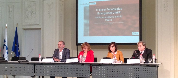
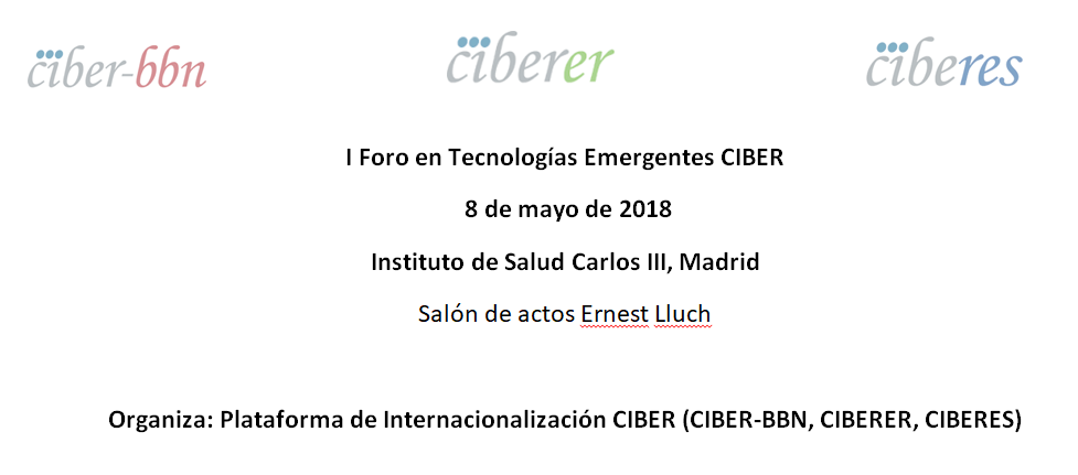
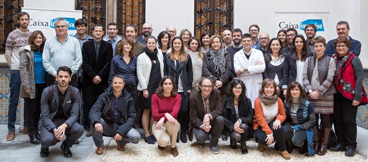
Nanotechnology, gene therapy, omics therapies and ‘big data’ were the topics discussed in the Forum on Emerging Technologies oganized on May 8 by nanotechnology, gene therapy, omics therapies and ‘big data’ were the topics discussed in the I Forum on Emerging Technologies held on May 8, organized by the CIBER Internationalization Platform, of which the CIBERER, the CIBER-BBN and the CIBERES.
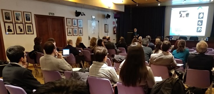
The objective of this event, structured in sessions of presentations and scientific debates about the types of technologies addressed, was to promote the exchange of ideas and scientific knowledge with the aim of generating new collaborations among the CIBER research groups such as participation in transversal projects or the development of cutting-edge technologies.
NANBIOSIS was represented by Pablo Laguna (Unit 27, High Performance Computing), Laura Lechuga (Unit 4, Biodeposition and Biodetection Unit), José Luis Pedraz (Unit 10, Drug Formulation) and Rosa Villa (Unit8, Micro – Nano Technology Unit)