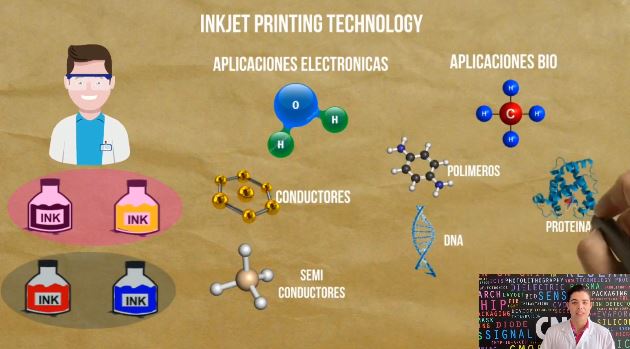
+34 9340006100 -ext. 437807-info@nanbiosis.com

News U8

Rosa Villa, Scientific Director of NANBIOSIS U8, New CIBER-BBN group leader

Rosa Villa Sanz, the Scientific Director of NANBIOSIS U8 Micro– Nano Technology Unit was appointed group leader the CIBER during the last meeting of the Permanent Commission of CIBER-BBN, replacing Prof. Jordi Aguiló.

Rosa Villa Sanz, graduated in Medicine from the University of Barcelona and has a doctorate from the Autonomous University of Barcelona in 1993 and a specialist in Nuclear Medicine (MIR-Hospital de Bellvitge-Barcelona), is a scientific researcher at the CSIC and leader of the Biomedical Applications Group of the Barcelona Institute of Microelectronics (CSIC), whose main research interests are the design and manufacture of Micro and Nano Systems for Biomedical Applications.

Prof. Villa has participated in more than 30 national and European projects, being a principal researcher in 10 of them, she has obtained the approvals by the Spanish Agency of Medicines and Medical Devices (AEMPS) for the conduct of 4 clinical trials in collaboration with pharmaceutical companies and clinical groups and is the founder of a spin-off (Barcelona Liver Bioservice SL). Rosa Villa also directs a consolidated research group recognized by the Agència de Gestió d’Ajuts Universitaris i de Recerca since 2014 (Bio-Micro-Nano-Systems group [2019 SGR 988]) and since 2016 co-directs the BioMEM TECNIO group of the Generalitat de Catalunya. Rosa Villa is Scientific of NANBIOSIS Unit 8 since the creation of NANBIOSIS.

Read More

Organ-on-chip monitoring. Breakthrough technological approximations

Organ-on-chip (OOC) is the term used to define a microfluidic 3D culture model that contains continuously perfused chambers inhabited by living cells. OOC are considered as very promising tools for investigating many aspects of human physiology and pathophysiology as well as drug testing platforms with future progressions to be used for precision medicine.

As the complexity of OOC systems increases, the necessity to integrate relevant assessment methods to provide information about cell physiology, secreted metabolites as well as pharmacodynamics drug responses also increases. Dr. Rosa Villa, who leads NANBIOSIS U8 Nano Technology Unit and the Biomedical Applications Group of the Institute of Microelectronics of Barcelona and CIBER in Bioengineering, Biomaterials and Nanomedicine, works on different engineering approaches to develop physical and chemical sensors that can be integrated into the OOC devices. The group considers that sensors integration is a requirement that must be taken into consideration in an OOC platform giving the necessary assessment of the OOC platforms in a continuous and real-time

An overview of the most relevant works of the Biomonitoring Group and NANBIOSIS Unit 8 have been presented by Mar Alvarez and Gemma Gabriel, researchers of
NANBIOSIS U8 Nano Technology in the conference on Engineering Multicellular Systems organized by EMBL – IBEC that took place in La Pedrera Auditorium, in Barcelona, from 10-12th February 2020.

It has been presented a device fabricated for that mimics Retina. In this novel microfluidic device cells are arranged in parallel compartments and are highly interconnected through a grid of microgrooves, which facilitates paracrine signaling and heterotypic cell–cell contact between multiple tissues. In the field of Brain, TEER barrier monitoring is mandatory. An interdigitated electrodes (IDE) configuration where the entire cell culture area contributes equally to the measurement, has been integrated in a custom-made bioreactor. This configuration, besides being more accurate for measuring the TEER, also allows the minimal electrode coverage, so that the optical visualization of the cell culture is maximized. The control and monitoring of dissolved oxygen (DO) is key for most of the OOC. The integration of oxygen sensors in an Liver-On-a-Chip system to achieve in-situ and real-time monitoring of oxygen zonation along the cell culture microfluidic chamber. A miniaturized sensing device compatible with microfluidic technology to measure simultaneously dissolved oxygen, pH, Na+ and K+, able to be connected in the input or output of a cell culture system has been developed for Kidney monitoring.

References

[1]   Yeste J, García-Ramírez M, Illa X, Guimerà A, Hernández C, Simó R, Villa R, “A compartmentalized microfluidic chip with crisscross microgrooves and electrophysiological electrodes for modeling the blood–retinal barrier” Lab on a Chip 18 (2018) 95-105

[2] Yeste J, Martínez-Gimeno L, Illa X, Laborda P, Guimerà A, Sánchez-Marín JP, Villa R, Giménez I “A perfusion chamber for monitoring transepithelial NaCl transport in an in vitro model of the renal tubule “, Biotechnology and Bioengineering 115 (2018) 1604-1613

[3] Moya A, Ortega-Ribera M, Guimerà X, Sowade E, Zea M, Illa X, Ramon E, Villa R, Gracia-Sancho J, Gabriel G., “Online oxygen monitoring using integrated inkjet-printed sensors in a Liver-On-a-Chip system” Lab on a Chip (2018),18, 2023-2035

[4]Moya A, Illa X, Gimenez I, Lazo-Fernandez Y, Villa R, Errachid A, Gabriel G. “Miniaturized multiparametric flexible platform for the simultaneous monitoring of ionic compounds: Application in real urine” Sensors and Actuators B: Chemical 255 (2018) 2861-2870

Read More

Researchers from NANBIOSIS Unit 8 opt for the Cutting-Edge Science Award: How to measure brain’s hidden activity.

La Vanguardia and the Fundació Catalunya La Pedrera have jointly promoted for the tenth consecutive year the Cutting-Edge Science Award “La Vanguardia de la Ciencia”, with the objective of givin visibility to the research of excellence carried out in Spain. The prize will correspond to the proposal candidate most voted by the public.

One of the 8 selected candidates is the research led by Anton Guimerà-Brunet (NANBIOSIS Unit 8 -Institut de Microelectrònica de Barcelona-CNM-CSIC / CIBER-BBN) and Jose Garrido (Institut Català de Nanociències i Nanotecnologia / Icrea), for developing graphene implants capable of measuring the hidden activity of the brain  with more sensitivity than conventional methods.

These new devices could improve the diagnosis of epilepsy and are being used as research tool to better understand this and other diseases and develop new therapies.

Further information can be found at the Vanguardias’s website dedicated to the prize and also how to cast your vote: https://www.lavanguardia.com/ciencia/20200126/473088817129/premio-vanguardia-de-la-ciencia.html

Article of reference:

Read More

Inkjet printing and paper sensors to control different analytes with low cost technologies

‘I investigate, I am CSIC’ is a competition hold by The Spanish National Research Council (CSIC) for its doctoral students to disseminate their doctoral thesis. Through short videos of maximum duration of 3 minutes, predoctoral scientists explain their research and results in an informative language.

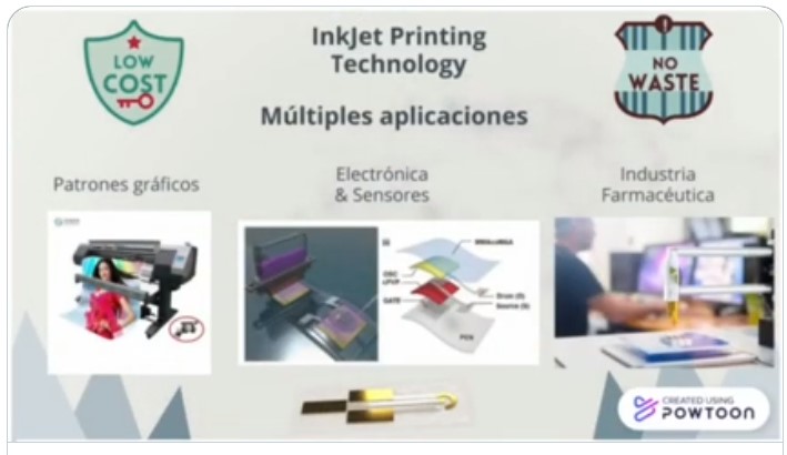
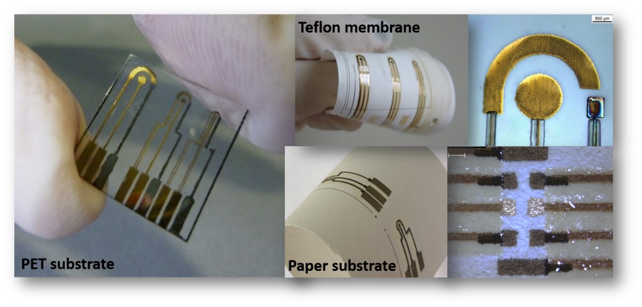
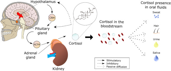
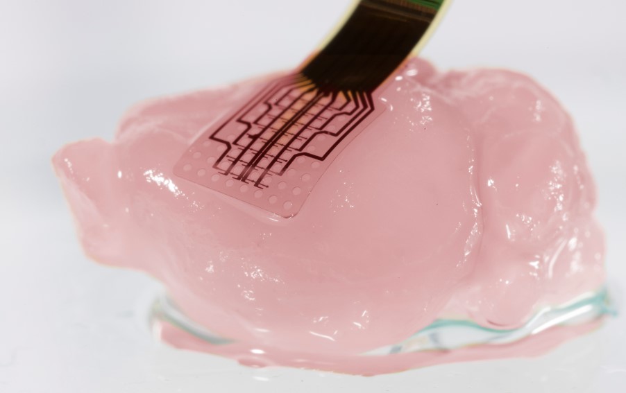
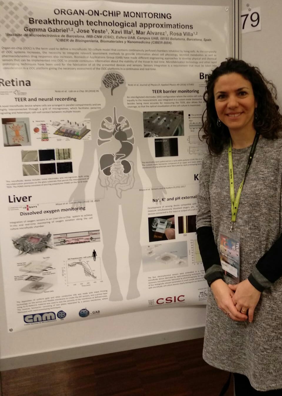
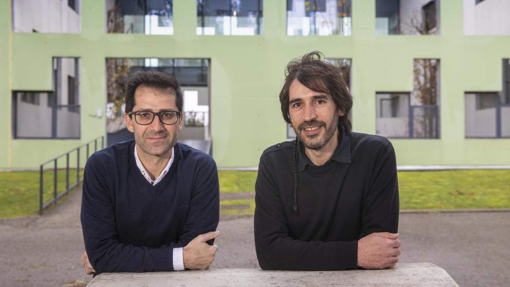
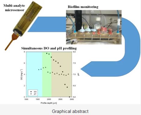
Miguel Zea, a member of the NANBIOSIS U8 Micro– Nano Technology Unit presents his video explaining how paper sensors can be manufactured to control different analytes with low cost technologies such as Inkjet Printing.

A jury composed of five experts in communication or scientific dissemination will choose eight videos taking into account the originality, impact, convenience and consistency of the video content. In addition, clarity will be valued when exposing the research work and the communicative capacity of the participant. The votes of the public through the YouTube channel of the Postgraduate Department will decide the selection of two other participants.

Read More

A Minimally Invasive Microsensor Specially Designed for Simultaneous Dissolved Oxygen and pH Biofilm Profiling

Ana Moya, Xavi Illa, Rosa Villa and Gemma Gabriel, researchers of the Biomedical Application’s Group (GAB) of CIBER-BBN and the Microelectronics Institute of Barcelona, which main interests are the design and fabrication of Micro and Nano Systems for Biomedical Applications, have recently published, in collaboration with other authors an article in the Special Issue Advanced Electrochemical Sensors and Environmental Monitoring

The authors thank in the Acknowledgements the participation of the ICTS NANBIOSIS U8 Micro– Nano Technology Unit of the CIBER in Bioengineering, Biomaterials and Nanomedicine (CIBER-BBN) at the IMB -CNM (CSIC)

The work reports the development of a novel multi-analyte microsensor based on MEMS technologies for simultaneous monitoring of DO and pH. Results showed the capacity of microsensors to quantify concentration gradients of di erent species within biofilms. In addition, the possibility of integrating two microsensors, specially designed to obtain a seven-point profile in a single measurement, introduced in this paper for the first time, clearly simplifies the equipment and the procedure necessary to record concentration profiles within biofilms. Critical future prospects have been established

Article:

Xavier Guimerà , Ana Moya, Antonio David Dorado, Xavi Illa, Rosa Villa, David Gabriel, Xavier Gamisans and Gemma Gabriel. A Minimally Invasive Microsensor Specially Designed for Simultaneous Dissolved Oxygen and pH Biofilm Profiling Sensors 201919(21), 747; https://doi.org/10.3390/s19214747

Read More

The hidden brain activity

Researchers of NANBIOSIS Unit 8 Micro and nano techonlogy unit, (Institut de Microelectrònica de Barcelona-CNM-CSIC/ CIBER-BBN ), led by Anton Guimera are coauthors of recently published article in the scientific magazine Nature Materials about the hidden brain activity, entitled “High-resolution mapping of infraslow cortical brain activity enabled by graphene microtransistors “

Recording infraslow brain signals (<0.1 Hz) with microelectrodes is severely hampered by current microelectrode materials, primarily due to limitations resulting from voltage drift and high electrode impedance. Hence, most recording systems include high-pass filters that solve saturation issues but come hand in hand with loss of physiological and pathological information. In this work, we use flexible epicortical and intracortical arrays of graphene solution-gated field-effect transistors (gSGFETs) to map cortical spreading depression in rats and demonstrate that gSGFETs are able to record, with high fidelity, infraslow signals together with signals in the typical local field potential bandwidth. The wide recording bandwidth results from the direct field-effect coupling of the active transistor, in contrast to standard passive electrodes, as well as from the electrochemical inertness of graphene. Taking advantage of such functionality, we envision broad applications of gSGFET technology for monitoring infraslow brain activity both in research and in the clinic.

Article of reference:

Masvidal-Codina, E., Illa, X., Dasilva, M. et al. High-resolution mapping of infraslow cortical brain activity enabled by graphene microtransistors. Nature Mater18, 280–288 (2019). https://doi.org/10.1038/s41563-018-0249-4

Read More

NANBIOSIS Scientific Women in the International Day of Women and Girls in Science

Today February 11 is the International Day of Women and Girls in Science, a day to raise awareness of the gender gap in science and technology.

According to the United Nations, while yet women and girls continue to be excluded from participating fully in science, science and gender equality are vital to achieve the internationally agreed development goals, including the 2030 Agenda for Sustainable Development. Thus, in recent years, the international community has made a great effort to inspire and promote the participation of women and girls in science.

NANBIOSIS wants to acknowledge  the efforts made by scientific women who struggle every day to contribute their bit to Science and highlight their essential role in nowadays research. Especially we want to recognize the work of scientists women involved in our units, whatever is the nature of their contribution: technical, scientific development, management, coordination, direction, etc; just to mention some examples:
Neus Ferrer in the Scientific Direction of Unit 1 Protein Production Platform (PPP)
Pilar Marco and Nuria Pascual in the Management and Scientific Coordination of U2 Custom Antibody Service (CAbS) 
Miriam Royo in the Scientific Direction of U3 Synthesis of Peptides Unit
Laura Lechuga and M.Carmen Estevez in the Direction and Scientific Coordination of U4 Biodeposition and Biodetection Unit
Nora Ventosa and Nathaly Segovia in the Scientific Direction and Technical Coordination of U6 Biomaterial Processing and Nanostructuring Unit
Isabel Oliveira and Teresa Galán in the Coordination of U7 Nanotecnology Unit
Rosa Villa and Gemma Gabriel in the Management and Scientific Coordination of U8 Micro – Nano Technology Unit
Gema Martínez in the Scientific Coordination of U9 Synthesis of Nanoparticles Unit
Fany Peña in the Scientific Coordination of U13 Tissue & Scaffold Characterization Unit
Mª Luisa González Martín in the of Direction and Scientific Coordination of U16 Tissue & Scaffold Characterization Unit
Gemma Pascual and Isabel Trabado in the Coordination of the U17 Confocal Microscopy Service
Mª Virtudes Céspedes in the Scientific Coordination of U18 Nanotoxicology Unit
Beatriz Moreno in the Scientific Direction of Unit 19 Clinical tests lab
Ibane Abásolo in the Scientific Coordination of Unit 20 In Vivo Experimental Platformt
Verónica Crisóstomo in the Scientific Direction of Unit 24 Medical Imaging 
Ana Paula Candiota in the Scientific Coordination of Unit 25 Biomedical Applications I 
Maria Luisa García in the Scientific Direction of U28 NanoImaging Unit from Bionand, recently incorporated to NANBIOSIS

Thanks to all of you and your teams!

Read More

Rosa Villa explains how New graphene implants can help to better understand the brain


A graphene implant that detects brain activity at extremely low frequencies could improve the technology of the electrodes to analyze the state of our brain, has been developed by researchers from several research institutes of the CSIC in Catalonia and the CIBER-BBN.

Last First of February , Rosa Villa, Scientific Director of NANBIOSIS U8 Micro – Nano Technology Unit was interviewed in Ágora, a program of Scientific Dissemination of Radio Aragón. Dr. Villa, researcher of the Biomedical Applications Group of the Institute of Microelectronics of Barcelona and CIBER in Bioengineering, Biomaterials and Nanomedicine, explains the relevance of the research carried out, together with several institutes of the CSIC in Catalonia, on the application of new materials to the study of brain activity.

The brain is composed of many neurons that communicate with each other. This communication occurs through electric currents that are detected with electrodes placed on the surface of the head or above the brain. Brain waves are very different if we are awake or asleep or when we have certain pathologies. The electrodes with which these electrical signals
were analyzed used to be large; thanks to the microelectronics began to make increasingly smaller electrodes that could identify communications much better but that small size also makes their limited reach, since they do not always take all the degrees of frequency.

Graphene has opened the degree of frequencies to detect the electrical signals of the brain. So far the electrodes were placed on top of the hair (for example the encephalograms) but now, although it has only been done in animals for the moment, the microelectrodes are already being placed as implants on the brain itself, which are left on the surface or they dig in to access more depth. When this is done, the brain feels invaded and isolates that electrode generating a scar, which is why more compatible materials are sought that are not rejected by the brain, such as graphene. Overcoming this technical limitation makes accessible the large amount of information that is below 0.1 Hz, while it facilitates the design of new brain-computer interfaces can register a wide range of frequency of what is occurring in a site of the brain.

Read More

Involvement of Cellular Prion Protein in α-Synuclein Transport in Neurons

Researchers of NANBIOSIS U7 Nanotechnology Unit are coauthors of the article “Involvement of Cellular Prion Protein in α-Synuclein Transport in Neurons” by Molecular Neurobiology

The cellular prion protein, encoded by the gene Prnp, has been reported to be a receptor of β-amyloid. Their interaction is mandatory for neurotoxic effects of β-amyloid oligomers. In this study, we aimed to explore whether the cellular prion protein participates in the spreading of α-synuclein. Results demonstrate that Prnp expression is not mandatory for α-synuclein spreading. However, although the pathological spreading of α-synuclein can take place in the absence of Prnp, α-synuclein expanded faster in PrPC-overexpressing mice. In addition, α-synuclein binds strongly on PrPC-expressing cells, suggesting a role in modulating the effect of α-synuclein fibrils.

Article: doi: 10.1007/s12035-017-0451-4


Read More

Resemblance of the human liver sinusoid in a fluidic device with biomedical and pharmaceutical applications

Scientists of Unit 8 of NANBIOSIS are co-authors of the article “Resemblance of the human liver sinusoid in a fluidic device with biomedical and pharmaceutical applications“, recently published  by Biotechnology and Bioengineering.  The fabrication of Exoliver was performed by the platform of Production of Biomaterials and Biomolecules of the ICTS, more specifically by the “NANBIOSIS,” Unit 8.

Maintenance of the complex phenotype of primary hepatocytes in vitro represents a limitation for developing liver support systems and reliable tools for biomedical research and drug screening. We herein aimed at developing a biosystem able to preserve human and rodent hepatocytes phenotype in vitro based on the main characteristics of the liver sinusoid: unique cellular architecture, endothelial biodynamic stimulation, and parenchymal zonation. Primary hepatocytes and liver sinusoidal endothelial cells (LSEC) were isolated from control and cirrhotic human or control rat livers and cultured in conventional in vitro platforms or within our liver‐resembling device. Hepatocytes phenotype, function, and response to hepatotoxic drugs were analyzed. Results evidenced that mimicking the in vivo sinusoidal environment within our biosystem, primary human and rat hepatocytes cocultured with functional LSEC maintained morphology and showed high albumin and urea production, enhanced cytochrome P450 family 3 subfamily A member 4 (CYP3A4) activity, and maintained expression of hepatocyte nuclear factor 4 alpha (hnf4α) and transporters, showing delayed hepatocyte dedifferentiation. In addition, differentiated hepatocytes cultured within this liver‐resembling device responded to acute treatment with known hepatotoxic drugs significantly different from those seen in conventional culture platforms. In conclusion, this study describes a new bioengineered device that mimics the human sinusoid in vitro, representing a novel method to study liver diseases and toxicology.

Article of reference: https://doi.org/10.1002/bit.26776

Read More